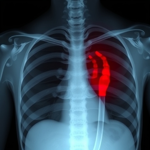
In a groundbreaking study published recently in Nature Communications, researchers have unveiled compelling evidence highlighting the predictive power of abnormal chest X-rays in foreseeing the development of tuberculosis (TB), a disease that remains a significant global health challenge. This pioneering investigation challenges existing paradigms in TB diagnosis and prognosis, emphasizing that subtle radiological anomalies can serve as critical harbingers of disease progression, thereby opening new frontiers in early intervention strategies.
Tuberculosis, caused by Mycobacterium tuberculosis, is notoriously difficult to detect in its early stages due to its often asymptomatic or nonspecific clinical presentation. Traditionally, diagnosis hinges on microbiological assays or the presence of overt clinical symptoms. However, this groundbreaking research shifts the spotlight towards radiological imaging, specifically chest X-rays, as an invaluable tool for predicting disease trajectory well before clinical manifestations become apparent.
The team, led by Zhu et al., systematically analyzed chest radiographs from a vast cohort of individuals at risk of tuberculosis infection. By meticulously correlating radiographic abnormalities with longitudinal health outcomes, the study demonstrated that an abnormal chest X-ray greatly increases the likelihood of progression to active TB within a defined period. This finding underscores the crucial prognostic value of radiographic screening, especially in high-burden settings.
Sophisticated image analysis techniques played a pivotal role in this research. Utilizing advanced computational methods and machine learning algorithms, the scientists were able to discern subtle radiological patterns that might elude even experienced radiologists. These patterns, often localized and non-specific, were nevertheless predictive markers of pathology, indicating latent or incipient infection stages.
This research fills a critical gap in the continuum of tuberculosis diagnosis. While sputum tests and microbiological cultures remain the gold standard for active disease confirmation, they offer limited utility in predicting future disease onset in asymptomatic individuals. The incorporation of chest X-ray abnormalities as prognostic indicators bridges this gap, providing a proactive lens through which clinicians can identify and monitor high-risk patients more effectively.
Another noteworthy aspect of the study lies in its epidemiological breadth. By encompassing diverse populations with varying TB exposure risks, the researchers ensured that their findings are robust and generalizable. This broad approach enables the adaptation of their screening protocols across different healthcare infrastructures, potentially revolutionizing how TB surveillance is conducted globally.
Moreover, the research outlines important clinical implications for TB control programs. Early identification of individuals with abnormal chest X-rays enables targeted preventive therapy, reducing the reservoir of latent infection and curtailing transmission chains. This strategic shift from reactive to proactive care could significantly impact disease incidence, particularly in regions with limited access to comprehensive diagnostic resources.
From a technical perspective, the study rigorously evaluated interobserver variability in chest X-ray interpretation, establishing that even minor abnormalities carry prognostic significance. The authors advocate for enhanced radiological training and standardized reporting protocols to maximize the clinical utility of radiographic data. This calls for integration of emerging technologies like artificial intelligence to augment human expertise in diagnostic accuracy.
The findings also bear relevance for public health policymakers, emphasizing resource allocation towards improving radiological infrastructure and training. Given the cost-effectiveness of chest X-rays relative to molecular tests, scaling up radiographic screening can be a feasible and impactful strategy in lower-resource settings, enabling earlier interventions and better outcomes.
The potential for leveraging this radiological prognostic marker extends beyond TB. The methodological framework developed by Zhu et al. could be adapted to other infectious or chronic lung diseases where early subtle imaging changes presage clinical deterioration, marking a new era in personalized radiology-driven healthcare.
Academic and clinical communities have lauded this research for its innovation and translational potential. The study’s robust statistical analyses and longitudinal design lend credibility and rigor, making it a seminal work that will likely influence future TB diagnosis guidelines and spark further research into radiology-based predictive diagnostics.
In summary, this study redefines the utility of chest X-rays from a mere diagnostic tool to a prognostic instrument with considerable predictive accuracy for tuberculosis development. By identifying abnormal radiological signals early, clinicians can implement timely interventions, ultimately reducing disease burden and transmission worldwide.
As tuberculosis continues to pose a formidable public health threat, particularly in under-resourced environments, innovations like those presented in this research provide hope for more effective management strategies. The integration of chest X-ray abnormalities into TB screening protocols could transform clinical practice and substantially accelerate global efforts to curb this devastating disease.
Future research stemming from this study could delve deeper into the mechanistic underpinnings of these radiological anomalies, potentially correlating imaging findings with immunological and microbiological markers to refine risk stratification further. Such multidimensional approaches promise to deepen understanding and heighten predictive capabilities for tuberculosis and beyond.
This work, therefore, represents a landmark advance, kindling new possibilities in the fight against tuberculosis and underscoring the enduring value of radiological imaging in infectious disease management. The adoption of these insights by healthcare systems worldwide could herald a new chapter in global health security.
Subject of Research: Prognostic value of abnormal chest X-rays in predicting tuberculosis development
Article Title: Prognostic value of an abnormal chest X-ray result in predicting the development of tuberculosis
Article References:
Zhu, P., Chen, X., Pan, J. et al. Prognostic value of an abnormal chest X-ray result in predicting the development of tuberculosis. Nat Commun 16, 9866 (2025). https://doi.org/10.1038/s41467-025-64834-9
Image Credits: AI Generated
DOI: https://doi.org/10.1038/s41467-025-64834-9
Tags: abnormal chest X-raysasymptomatic tuberculosis diagnosischest radiograph analysisearly TB intervention strategieshealth outcomes correlationhigh-burden TB settingslongitudinal health studiesMycobacterium tuberculosis detectionNature Communications research findingsprognostic value of radiographic screeningradiological imaging in TBtuberculosis risk prediction


