• HOME
  • NEWS
  • EXPLORE
    • CAREER
      • Companies
      • Jobs
    • EVENTS
    • iGEM
      • News
      • Team
    • PHOTOS
    • VIDEO
    • WIKI
  • BLOG
  • COMMUNITY
    • FACEBOOK
    • INSTAGRAM
    • TWITTER
Monday, April 13, 2026
BIOENGINEER.ORG
No Result
View All Result
  • Login
  • HOME
  • NEWS
  • EXPLORE
    • CAREER
      • Companies
      • Jobs
        • Lecturer
        • PhD Studentship
        • Postdoc
        • Research Assistant
    • EVENTS
    • iGEM
      • News
      • Team
    • PHOTOS
    • VIDEO
    • WIKI
  • BLOG
  • COMMUNITY
    • FACEBOOK
    • INSTAGRAM
    • TWITTER
No Result
View All Result
Bioengineer.org
No Result
View All Result
Home NEWS Science News Biology

Super-resolution microscopy builds multicolor 3D from 2D

Bioengineer by Bioengineer
October 1, 2018
in Biology
Reading Time: 2 mins read
0
Share on FacebookShare on TwitterShare on LinkedinShare on RedditShare on Telegram
IMAGE

Credit: Christian Sieben/EPFL

Super-resolution microscopy is a technique that can "see" beyond the diffraction limit of light. The technique has garnered increasing interest recently, especially since its developers won the Nobel Prize in Chemistry in 2014. By exploiting fluorescence, super-resolution microscopy now allows scientists to observe cells and their interior structures and organelles in a way never before possible.

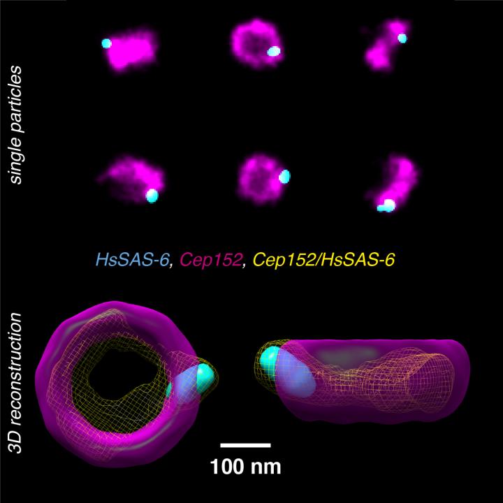
Many of the molecular complexes inside cells are made up of multiple proteins. Since current techniques of super-resolution microscopy typically can only use one or two fluorescent colors, it is difficult to observe different proteins and decipher the complex architecture and underlying assembly mechanisms of the cell's interior structures. An even greater challenge is to overcome the noise inherent to the super-resolution methods and fluorescent labeling, to achieve the full resolution potential.

Scientists from the lab of Suliana Manley at EPFL have now solved both problems by developing a new method to analyze and reconstruct super-resolution images and re-align them in a way that multiple proteins can be placed within a single 3D volume. The method works with images taken with large field-of-view super-resolution microscopy, with each image containing hundreds of two-dimensional projections of a labeled structure in parallel.

Each 2D view represents a slightly different orientation of the structure, so that with a dataset of thousands of views, the method can computationally reconstruct and align the 2D images into a 3D volume. By combining information from a large number of single images, the noise is reduced and the effective resolution of the 3D reconstruction is enhanced.

With the help of Pierre Gönczy's lab at EPFL, the researchers tested the method on human centriole complexes. Centrioles are pairs of cylindrical molecular assemblies that are crucial in helping the cell divide. Using the new multicolor super-resolution reconstruction method, the researchers were able to uncover the 3D architecture of four proteins critical for centriolar assembly during organelle biogenesis.

The new approach allows for unlimited multiplexing capabilities. "With this method, if the proteins in the structure can be labeled, there is no limit to the number of colors in the 3D reconstruction," says Suliana Manley. "Plus, the reconstruction is independent of the super-resolution method used, so we expect this analysis method and software to be of broad interest."

###

Reference

Christian Sieben, Niccolò Banterle, Kyle M. Douglass, Pierre Gönczy, Suliana Manley. Multicolor single particle reconstruction of protein complexes. Nature Methods 01 October 2018. DOI: 10.1038/s41592-018-0140-x

Media Contact

Nik Papageorgiou
[email protected]
41-216-932-105
@EPFL_en

http://www.epfl.ch/index.en.html

Related Journal Article

http://dx.doi.org/10.1038/s41592-018-0140-x

Share12Tweet8Share2ShareShareShare2

Related Posts

WCM Investigators Harness AI to Empower Cancer Research

WCM Investigators Harness AI to Empower Cancer Research

April 13, 2026
Tracing Arabidopsis Development Reveals Three-Cell Branching Rule

Tracing Arabidopsis Development Reveals Three-Cell Branching Rule

April 13, 2026

Intensive Control Transforms Malaria Genetics in Southeast Asia

April 13, 2026

Innovative Light-Activated Mechanism Unveiled for Regulating Plant Growth

April 13, 2026
Please login to join discussion

POPULAR NEWS

  • Scientists Investigate Possible Connection Between COVID-19 and Increased Lung Cancer Risk

    59 shares
    Share 24 Tweet 15
  • Boosting Breast Cancer Risk Prediction with Genetics

    47 shares
    Share 19 Tweet 12
  • Popular Anti-Aging Compound Linked to Damage in Corpus Callosum, Study Finds

    45 shares
    Share 18 Tweet 11
  • Imagine a Social Media Feed That Challenges Your Views Instead of Reinforcing Them

    1012 shares
    Share 400 Tweet 250

About

We bring you the latest biotechnology news from best research centers and universities around the world. Check our website.

Follow us

Recent News

Gut Microbiota Biomarkers Predict Rectal Cancer Therapy Response

Fungal Endophytes in Cacti Could Boost Cacao’s Drought Resilience

High-Resolution Long-Read Sequencing Reveals Myotonic Dystrophy Repeats

Subscribe to Blog via Email

Enter your email address to subscribe to this blog and receive notifications of new posts by email.

Join 79 other subscribers
  • Contact Us

Bioengineer.org © Copyright 2023 All Rights Reserved.

Welcome Back!

Login to your account below

Forgotten Password?

Retrieve your password

Please enter your username or email address to reset your password.

Log In
No Result
View All Result
  • Homepages
    • Home Page 1
    • Home Page 2
  • News
  • National
  • Business
  • Health
  • Lifestyle
  • Science

Bioengineer.org © Copyright 2023 All Rights Reserved.