Credit: © NUST MISIS
A research team from NUST MISIS and Pirogov Russian National Research Medical University (RNRMU) has successfully conducted preclinical trials of a new anticancer drug based on magnetite nanoparticles. In the test results, the lifetime of sick mice who were given the drug was doubled. The research results have been published in Nanomedicine.
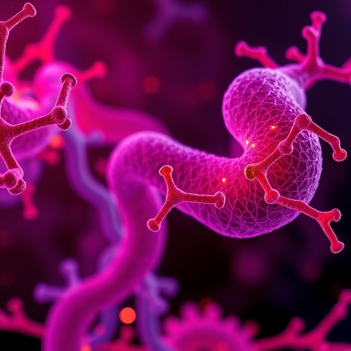
This innovative drug is a combination of two components: spherical nanoparticles of magnetite, which contain a cytostatic drug (a toxic substance that destroys tumor cells), and a vector molecule that delivers the drug to its destination. Essentially, the molecule leads the poisoned particle specifically to the affected organ, ensuring that healthy tissues are not exposed.
«NUST MISIS scientists, led by Maxim Abakumov, Candidate of Technical Sciences and Head of the NUST MISIS Biomedical Nanomaterials Laboratory, have been working with magnetite nanoparticles for four straight years, and they're [now] using them to create anticancer drugs. Currently, the research team is working on the preparation of the preclinical trials stage, which are planned for 2019», said Alevtina Chernikova, Rector of NUST MISIS.
The vector molecule is an antibody to the protein of the «vascular endothelial growth factor» – a signaling protein produced by cells to stimulate the formation of the embryonic vascular system. The molecule works as a «key-lock», finding and joining only to certain types of cells. In this study, the scientists were among the first in the world to use a vector molecule in such an unusual function, considering its use historically as an independent drug. However, monotherapy using these antibodies has not shown high efficiency. Nevertheless, this doesn't make the protein less promising as an «address» for drug delivery, which has been demonstrated in the current work.
«The studies have shown that the proposed therapeutic scheme is effective: in vitro experiments have demonstrated that the animals` life expectancy when treated with the new drug increased by 50%, from 23 to 39 days. In addition to that, the proposed substance showed a good visualization of tumor tissue during MRI examination. This can potentially be used to facilitate the work of surgeons during surgery for marking and fixing the edges of affected organs», said Maxim Abakumov, head of the project and head of the NUST MISIS Biomedical Nanomaterials Laboratory.
In addition to the drug delivery scheme – which includes the current study – the iron oxide particles show good results as a method of therapy by hyperthermia.
The process is outlined as follows: the magnetite nanoparticles are introduced into the affected organ, where they accumulate and then are exposed to an alternating electromagnetic field with certain parameters; as a result of this exposure the nanoparticles are heated to 43-45 degrees and subsequently heat the surrounding cancer cells, causing them to die.
It is important to note that previous research has shown that cancer cells are more sensitive to temperature changes than healthy ones, which leaves open the opportunity to leave healthy tissue intact.
###
Media Contact
Lyudmila Dozhdikova
[email protected]
7-495-647-2309
http://en.misis.ru/
Original Source
http://en.misis.ru/university/news/science/2018-05/5391/ http://dx.doi.org/10.1016/j.nano.2018.04.019


