
Credit: Pauline Zulueta/Cumming School of Medicine
It's one of the most talked about injuries in sport today, concussion. Yet, there is no accepted way to image a concussion. University of Calgary scientist Jeff Dunn, PhD, hopes to change that. He and his team have developed a portable brain imaging system that uses light to detect and monitor damage in the brain from concussion. Researchers and doctors will be using the technology in an upcoming study at the Alberta Children's Hospital.
The device, a Near-Infrared Spectroscopy (fNIRS), measures communication in the brain by measuring oxygen levels. When the brain is working well, major regions on each side of the brain are communicating and so have similar patterns of blood flow and oxygen levels in blood. Researchers measure the changes in blood oxygen levels as a marker of brain function. Results show these patterns change after concussion.
"The one thing we know is that there can be physiological changes in the brain that last for months to years after a brain injury due to concussion. We discovered a new technology that is portable to measure those changes," says Dunn, director of the Experimental Imaging Centre at the Cumming School of Medicine (CSM). "We will now apply this new technology to see how important these changes are, as we try to understand how concussion evolves over time."
Dunn became interested in finding a way to image concussion after his children became involved in ski racing.
"A lot of kids on the ski hill were getting head hits and it was really very apparent that there wasn't an imaging method that could help them," says Dunn who is also a member of the Hotchkiss Brain Institute and Alberta Children's Hospital Research Institute at the CSM. "Currently, concussion is often measured by the symptoms someone experiences, but we really don't know what's happening in the brain in any one person."
Symptoms can vary greatly between individuals, and can include headaches, nausea, loss of memory and lack of coordination (to name a few), making it even more difficult to find treatment options for each person. Through his research, Dunn hopes the images will show a connection between symptoms and abnormalities in the brain that could help doctors identify treatment protocols and recovery timelines.
In partnership with Alberta Health Services and with the support of the Canadian Institutes of Health Research, Dunn will be teaming up with physicians at the Alberta Children's Hospital, and other UCalgary researchers at the CSM and in the faculties of Kinesiology and Arts to use the imaging device to study concussion in children.
"We will be able to follow young patients over time to establish whether this new technology can help us determine the extent of the injury and how the brain is recovering," says Dunn.
Keith Yeates, PhD, lead for the university's Integrated Concussion Research Program (ICRP) and a co-investigator on the study, says the technology has important practical benefits, "The fNIRS device has the potential to provide a convenient method for helping detect concussion."
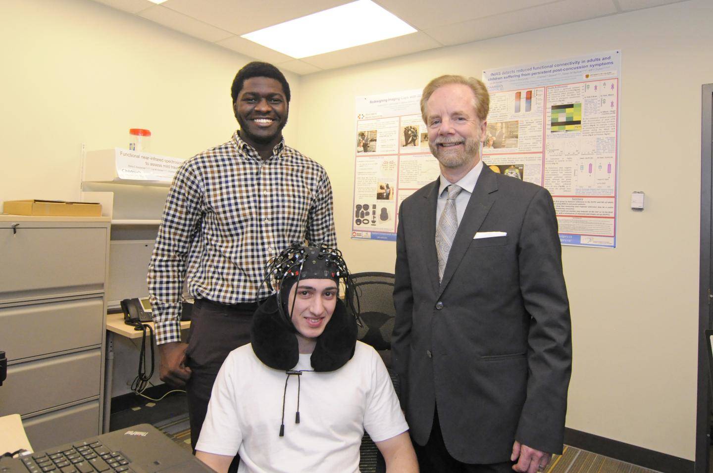
To image the brain, the researchers place a cap, similar to a swim or bathing cap, on the top of the head. The cap contains small lights that have sensors connected to a computer. When researchers turn on the lights, they can monitor and measure brain activity. The device is non-invasive and portable.
While most often associated with sports, concussion affects thousands of Canadians a year who experience a fall, or a car accident. While most people recover, some experience long-term effects and disabilities.
A recent study, by Dunn and colleagues, showed promising results of the device's ability to detect changes in brain activity in adults with long-term symptoms post-concussion. The findings are published in the Journal of Neurotrauma. This research is still ongoing at the Foothills Medical Centre.
If this new technology proves to be an effective tool for monitoring injury and recovery timelines Dunn hopes, one day, to see the technology widely used in concussion treatment clinics and sports facilities.
###
Led by the Hotchkiss Brain Institute, Brain and Mental Health is one of six research strategies guiding the University of Calgary towards its Eyes High goals. The strategy provides a unifying direction for brain and mental health research at the university and positions researchers to unlock new discoveries and treatments for brain health in our community.
The Hotchkiss Brain Institute and the Alberta Children's Hospital Research Institute are both partnerships between the University of Calgary's Cumming School of Medicine and Alberta Health Services.
Media Contact
Kelly Johnston
[email protected]
403-220-5012
@UCalgary
http://www.ucalgary.ca
Original Source
http://www.ucalgary.ca/utoday/issue/2018-03-15/ucalgary-researchers-develop-portable-brain-imaging-system-could-shed-light http://dx.doi.org/10.1089/neu.2017.5365


