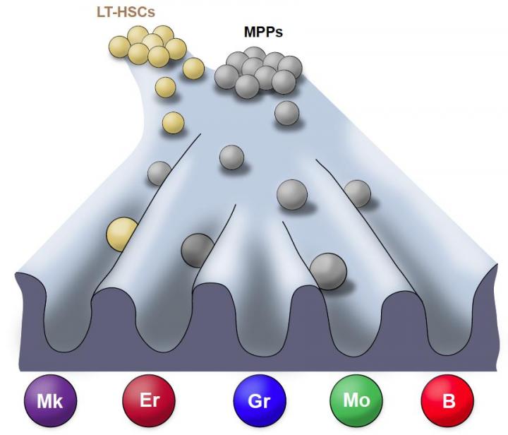
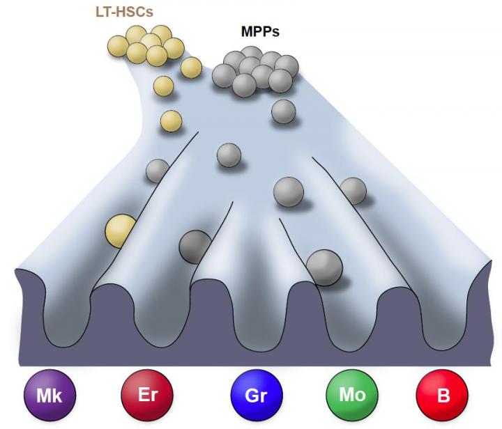
Credit: Courtesy of the Stem Cell Program, Boston Children's Hospital.
By tagging bone marrow cells of mice with a genetic label, or barcode, researchers were able to track and describe the family tree of individual blood cells as they form in their natural environment. The scientists discovered that these cells regenerate differently than their counterparts do after a blood cell transplant, according to a study published Jan. 3 in Nature and funded by the National Heart, Lung and Blood Institute (NHLBI), part of the National Institutes of Health.
"The findings of this research, if applicable to humans, will have implications for blood cell transplantation, and for clinical and research methods using blood cells, such as gene therapy or gene editing," said John W. Thomas, Ph.D., Stem Cell and Cell-based Therapy Coordinator at NHLBI.
This study moves research a step further towards the development of blood regeneration therapies, but the researchers believe it is also applicable to a variety of cells and will yield insights about regenerating diseased or damaged tissues.
"Our results show that stem cells and their less pluripotent descendants, blood progenitors, behave somewhat differently when studied without removing them from their native environment versus when studied in a laboratory or in transplantation; leading to differences in the type of blood lineages they make," said study's first author Alejo Rodriguez Fraticelli, Ph.D., from Harvard Stem Cell Institute, at the Boston Children's Hospital.
Due to the lack of appropriate tools to study how blood forms in the natural environment of the body, the majority of studies about where individual blood cells come from have been done after a transplant. In that context, the transplanted cells would have been "perturbed," or removed from their natural environment. According to the researchers, the current models are more likely to represent a roadmap of lineage potential for the blood cells' natural offspring.
For Rodriguez Fraticelli, this highlights the importance of studying blood regeneration in its native context. "Moving forward, we need to come up with methods to better predict what types of cells will be the most optimal for therapy, for instance in reprogramming cells, and editing," he said.
In the study, researchers tagged cells using a transposon, a piece of genetic code that can jump to a random point in DNA when exposed to an enzyme called transposase, to track blood progenitors and adult stem cells during the natural, unperturbed process of blood regeneration.
The research provides evidence for a substantially revised roadmap for normal blood regeneration or blood production in the natural environment; and highlights how in those conditions blood stem cells and progenitor cells manifest unique properties.
###
Part of the National Institutes of Health, the National Heart, Lung, and Blood Institute (NHLBI) plans, conducts, and supports research related to the causes, prevention, diagnosis, and treatment of heart, blood vessel, lung, and blood diseases; and sleep disorders. The Institute also administers national health education campaigns on women and heart disease, healthy weight for children, and other topics. NHLBI press releases and other materials are available online at http://www.nhlbi.nih.gov.
About the National Institutes of Health (NIH): NIH, the nation's medical research agency, includes 27 Institutes and Centers and is a component of the U.S. Department of Health and Human Services. NIH is the primary federal agency conducting and supporting basic, clinical, and translational medical research, and is investigating the causes, treatments, and cures for both common and rare diseases. For more information about NIH and its programs, visit http://www.nih.gov.
NIH…Turning Discovery Into Health
Media Contact
NHLBI Engagement and Media Relations Branch
[email protected]
301-496-5449
http://www.nhlbi.nih.gov
Original Source
https://www.nhlbi.nih.gov/news/2018/cellular-barcoding-helps-scientists-understand-behavior-stem-cells