Researchers investigating the molecular switches of systemic inflammation have uncovered an unexpected enzymatic mechanism that licenses the activation of the NLRP3 inflammasome, a multi-protein immune complex responsible for fueling severe inflammatory disorders such as sepsis. This newly identified protein assembly catalyzes an atypical ubiquitination reaction on the acetyltransferase KAT5, attaching ubiquitin through an unconventional oxyester linkage at serine 123. Under physiological and endotoxic conditions, this serine-directed modification substantially enhances KAT5 acetyltransferase activity, driving the robust acetylation and oligomeric assembly of NLRP3. When investigators disrupted this regulatory cascade in cellular systems and preclinical mouse models of lipopolysaccharide-induced endotoxemia, the hyperinflammatory cascade was largely suppressed, leading to reduced tissue destruction in vital organs and significantly enhanced animal survival.
The NLRP3 inflammasome operates as an intracellular sensor within myeloid cells, detecting pathogen-associated and damage-associated molecular patterns to trigger the maturation and secretion of potent pro-inflammatory cytokines such as interleukin-1beta. Canonical activation of this inflammasome proceeds through a two-stage sequence comprising priming and assembly. During priming, toll-like receptor engagement prompts transcriptional upregulation of inflammasome components, while secondary stimuli such as potassium efflux, extracellular adenosine triphosphate, or bacterial pore-forming toxins trigger complex assembly and caspase-1 activation. Previous research had highlighted post-translational modifications, including phosphorylation and acetylation, as pivotal regulators of inflammasome assembly. In particular, KAT5 had been established as an essential acetyltransferase that acetylates NLRP3 to permit its full oligomerization. However, the precise upstream molecular events governing the enzymatic activation of KAT5 itself had remained an unresolved mystery in cellular immunology, presenting a barrier to understanding how acetyltransferase activity is selectively unleashed during acute innate immune responses.
To investigate the upstream factors modulating this pathway, the researchers focused on DDB1- and Cul4-associated factor 15, known as DCAF15, an established substrate receptor for the CRL4 cullin-RING ubiquitin ligase family previously studied in oncological contexts and bacterial meningitis. Utilizing myeloid-specific conditional knockout mice, designated as Dcaf15cKO, the investigators harvested primary peritoneal macrophages to evaluate cellular responses following lipopolysaccharide challenge. Peritoneal macrophages lacking DCAF15 exhibited significant impairments in downstream inflammatory signaling cascades, displaying suppressed phosphorylation of IKK alpha/beta, NF-kappaB p65, and the MAP kinase ERK relative to wild-type controls. Genetic silencing of DCAF15 via small interfering RNA in primary macrophages corroborated these findings, yielding corresponding reductions in the secretion of tumor necrosis factor-alpha and interleukin-6. Furthermore, upon sequential stimulation with lipopolysaccharide and nigericin, Dcaf15-deficient macrophages failed to mount normal inflammasome responses, showing markedly decreased production of cleaved caspase-1 and mature interleukin-1beta along with diminished ASC speck formation.
The therapeutic significance of these cellular observations was demonstrated in vivo using mouse models subjected to systemic endotoxemia induced by intraperitoneal lipopolysaccharide injection. When monitored over a seven-day observation period following a lethal lipopolysaccharide challenge of 40 micrograms per gram of body weight, Dcaf15 conditional knockout mice exhibited a statistically significant survival advantage over wild-type counterparts, with a Mantel-Cox log-rank comparison yielding a significant protective divergence. In parallel cohorts administered a sublethal endotoxin dose of 25 micrograms per gram, serum interleukin-1beta concentrations were substantially lower in knockout animals after four hours of exposure. Histopathological evaluation of organ specimens stained with hematoxylin and eosin revealed that genetic ablation of DCAF15 protected against acute lung injury and splenic pathology. Knockout animals displayed reduced neutrophil infiltration, preservation of alveolar microarchitecture, diminished white pulp atrophy, and decreased extramedullary hematopoiesis, confirming that DCAF15 actively drives pathological inflammation and tissue injury in whole-animal endotoxemia.
Seeking the direct molecular target of DCAF15, the authors explored physical associations between DCAF15 and KAT5 through comprehensive biochemical and structural assays. Co-immunoprecipitation experiments performed in HEK293T cells expressing tagged proteins confirmed a robust interaction between DCAF15 and KAT5, while endogenous complexes were verified in mouse peritoneal macrophages following lipopolysaccharide stimulation. Subcellular imaging via confocal microscopy showed that both proteins predominantly localize to the nucleus in resting cells but partially redistribute toward the cytoplasm upon inflammatory stimulation in human THP-1 monocytes. In vitro GST pull-down experiments using recombinant proteins purified from Escherichia coli proved that the interaction is direct and independent of intermediate cellular bridging factors. Protein truncation mapping identified that amino acid residues 386 to 598 within DCAF15 specifically bind to the N-terminal region spanning residues 1 to 142 of KAT5, while rigid-body protein docking modeling predicted a stable binding interface with an estimated low binding energy of minus 16.8 kilocalories per mole stabilized by specific hydrogen bonding and salt bridge interactions.
Because DCAF15 functions as an E3 ligase adaptor, the authors investigated whether KAT5 represents a direct substrate for ubiquitination. Overexpression experiments in HEK293T cells and endogenous pulldown assays in primary peritoneal macrophages demonstrated that DCAF15 markedly increases KAT5 polyubiquitination, a modification that increased in a time-dependent manner following lipopolysaccharide exposure. Employing ubiquitin linkage mutants, the authors discovered that DCAF15 uniquely promoted atypical Lys6-linked polyubiquitination rather than classic Lys48 or Lys63 chains, a finding verified by linkage-specific immunoblotting. High-resolution liquid chromatography-tandem mass spectrometry of purified KAT5 identified four candidate ubiquitination sites: serine 123, serine 131, serine 188, and serine 195. Systematic site-directed mutagenesis replacing each serine with alanine revealed that only the KAT5 S123A substitution dramatically abolished DCAF15-dependent ubiquitination. Importantly, when all lysine residues in KAT5 were mutated to arginine in a lysine-less mutant, robust ubiquitination persisted in the presence of DCAF15, whereas combining the lysine-less mutation with the S123A substitution eliminated the signal entirely, providing strong evidence for non-canonical serine ubiquitination.
Because serine ubiquitination occurs through ester linkages rather than standard isopeptide bonds formed on lysine residues, the investigators subjected the modified proteins to chemical cleavage assays using strong nucleophiles. Treatment of immunoprecipitated complexes with one molar hydroxylamine or sixty millimolar sodium hydroxide completely cleaved and abrogated the polyubiquitin signal on KAT5, confirming the presence of chemically labile oxyester bonds. Interestingly, molecular dynamics simulations over a one-hundred-nanosecond production trajectory revealed that the KAT5 S123A mutant exhibited lower root mean square deviation and root mean square fluctuation values than the wild-type protein, reflecting an increase in overall conformational rigidity. These biophysical simulations indicated that wild-type serine 123 ubiquitination dynamically alters the structural flexibility of KAT5 without targeting the enzyme for 26S proteasomal degradation, distinguishing this regulatory modification from classical degradative ubiquitin cascades.
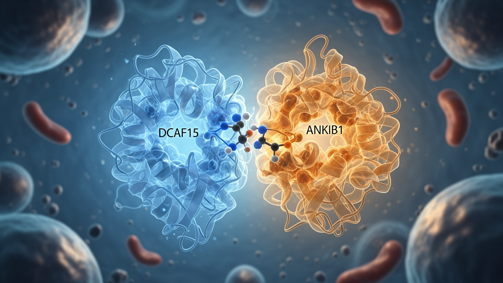
To resolve how DCAF15 catalyzes serine-directed ubiquitination despite lacking an intrinsic catalytic domain capable of ester bond formation, the researchers screened for interacting catalytic partners using tandem mass spectrometry in Flag-DCAF15-expressing THP-1 monocytes. Among the candidate interactors, the authors identified ANKIB1, an E3 ubiquitin ligase belonging to the RING-between-RING family known for mediating atypical ubiquitin linkages. Computational docking indicated a strong interface between DCAF15 and ANKIB1 characterized by a calculated binding energy of minus 57.56 kilocalories per mole, and physical interactions among endogenous proteins were confirmed by co-immunoprecipitation and confocal microscopy. Importantly, small interfering RNA knockdown of DCAF15 severely disrupted the binding of ANKIB1 to KAT5, demonstrating that DCAF15 serves as an obligatory scaffold facilitating complex assembly. This structural arrangement brings the substrate-binding module of CRL4-DCAF15 together with ANKIB1 into an integrated E3-E3 super-complex dedicated to orchestrating downstream substrate modification.
The functional competence of this enzymatic super-complex was reconstituted in an in vitro cell-free system using purified recombinant human E1 UBE1, E2 UbcH5c, ubiquitin, purified His-ANKIB1, reconstituted DCAF15 complex, and recombinant KAT5 in the presence of magnesium-ATP. Robust ubiquitination of wild-type KAT5 was observed in this defined cell-free environment, which was sensitive to cleavage by hydroxylamine and sodium hydroxide but was profoundly diminished when using the KAT5 S123A mutant. In cellular experiments, overexpression of wild-type ANKIB1 enhanced KAT5 ubiquitination, whereas ANKIB1 knockdown or the expression of a catalytic mutant carrying multiple cysteine-to-serine mutations across its RING2 domain significantly impaired ubiquitination and reduced NLRP3 acetylation. In cell lines stably expressing these constructs, the ANKIB1 RING2 mutant diminished caspase-1 activation and interleukin-1beta maturation, establishing that the catalytic integrity of ANKIB1 within the super-complex is required to license downstream KAT5-dependent inflammasome activation.
To evaluate the functional consequence of the Ser123 modification in vivo, the researchers employed recombinant adeno-associated viruses expressing either wild-type KAT5 or the ubiquitination-deficient KAT5 S123A mutant under the control of the macrophage-specific F4/80 promoter. Male C57BL/6J mice were administered these viral vectors four weeks prior to an intraperitoneal lipopolysaccharide challenge. Following endotoxin administration, animals expressing the KAT5 S123A mutant demonstrated remarkable protection from inflammatory pathology compared to mice transduced with wild-type KAT5. Similarly, spleen specimens from S123A-expressing mice exhibited preserved architecture, reduced sinusoidal congestion, and attenuated lymphocyte depletion. Quantitative enzyme-linked immunosorbent assays demonstrated that concentrations of interleukin-1beta were significantly decreased in both serum and bronchoalveolar lavage fluid from S123A animals, confirming that preventing Ser123 modification curtails systemic and pulmonary inflammatory pathology in intact organisms.
The investigators also observed a notable biological decoupling between cytokine release and cell death in cells expressing the KAT5 S123A mutant. The authors explained that interleukin-1beta maturation requires complete NLRP3 assembly, caspase-1 cleavage, and gasdermin D pore formation, whereas lytic cell death and lactate dehydrogenase release can be driven by parallel pathways, including non-canonical caspase-11 activation or NINJ1-dependent plasma membrane rupture. While these findings delineate a comprehensive regulatory axis, the authors documented several study limitations. The current data demonstrate that Ser123 is required for K6-linked polyubiquitination, but direct chemical identification of the branched ubiquitin chain on serine 123 awaits confirmation by high-resolution mass spectrometry. Additionally, because DCAF15 deletion impairs NF-kappaB and MAP kinase priming pathways, a partial contribution of decreased baseline component abundance cannot be completely decoupled from assembly defects. Nonetheless, the characterization of the DCAF15-ANKIB1-KAT5 axis reveals an unexpected post-translational control mechanism that may inform therapeutic strategies targeting excessive inflammasome activation in acute inflammatory disease.
Subject of Research: Regulation of NLRP3 inflammasome activation by the DCAF15-ANKIB1 ubiquitin ligase complex via KAT5 serine ubiquitination
Article Title: DCAF15-ANKIB1 E3 super-complex licenses NLRP3 inflammasome activation via KAT5 serine ubiquitination
Article References: Yin, H., Li, Y., Meng, A., Cao, X., Lang, S., Wang, H., Yan, R., Tian, Y., Liu, X., Li, Z., Peng, L., & Fang, R. (2026). DCAF15-ANKIB1 E3 super-complex licenses NLRP3 inflammasome activation via KAT5 serine ubiquitination. EMBO Reports. https://doi.org/10.1038/s44319-026-00987-y
Image Credits: AI Generated
DOI: 10.1038/s44319-026-00987-y
Keywords: NLRP3 inflammasome, DCAF15, ANKIB1, KAT5, serine ubiquitination, oxyester linkage, E3 ubiquitin ligase, cullin-RING ligase, RBR E3 ligase, interleukin-1beta, sepsis, endotoxemia, post-translational modification, protein acetylation, macrophages, caspase-1, ASC specks, inflammation
News Source: Drew Townsend. (November 22, 2026). DCAF15 and ANKIB1 Form Enzyme Complex Driving Inflammatory NLRP3 Activation. Scienmag.