A retrospective investigation examining 110 patients diagnosed with renal tuberculosis has elucidated the multifaceted clinical and pathological mechanisms responsible for progressive renal function decline, demonstrating that persistent mycobacterial infection of the renal parenchyma represents the primary and most direct cause of irreversible functional loss. Published in Frontiers in Medicine by a clinical research team from Hebei Chest Hospital and The Second Hospital of Hebei Medical University, the study analyzed medical records spanning from October 2013 to June 2024. The investigators tracked how renal deterioration unfolds across intrarenal vascular, renal parenchymal, and post-renal dimensions, revealing that severe functional impairment frequently proceeds even when patients receive standard medical therapy. By combining split-function nuclear scintigraphy, computed tomography, histopathological staining, and transmission electron microscopy, the authors documented that renal functional deterioration arises not merely from mechanical urinary obstruction, but from a coordinated triad of structural injuries including vascular narrowing, extensive parenchymal inflammation, and undetected contralateral excretory delays.
Genitourinary tuberculosis represents one of the most frequent manifestations of extrapulmonary tuberculosis, ranking second only to lymph node involvement and accounting for approximately 30 percent to 40 percent of all extrapulmonary cases. Clinical data cited by the authors indicate that roughly 4.8 percent of pulmonary tuberculosis infections are complicated by secondary renal seeding. Because Mycobacterium tuberculosis can spread down the urinary tract to induce secondary lesions in the ureters, urinary bladder, and urethra, the kidney occupies a central position in the therapeutic management of genitourinary tuberculosis. Even among individuals diagnosed relatively early who undergo standardized antituberculosis chemotherapy alongside ureteral stent drainage for hydronephrosis, progressive functional impairment often persists, culminating in permanent renal nonfunction and requiring surgical nephrectomy.
To systematically characterize the determinants driving this progressive loss of function, the investigators conducted a single-center retrospective analysis of 110 patients who had a definitive diagnosis of renal tuberculosis confirmed through urine Mycobacterium tuberculosis culture, tuberculosis DNA detection, or postoperative histopathology. The study population comprised 46 male patients (41.82 percent) and 64 female patients (58.18 percent), with an overall mean age of 44.88 years (standard deviation of 12.78 years, range 18 to 71 years). Unilateral renal tuberculosis accounted for 105 cases (95.45 percent), with 54 patients affected on the left side (49.09 percent) and 51 affected on the right side (46.36 percent). Only 5 patients (4.55 percent) presented with bilateral renal involvement. Strict exclusion criteria were enforced to isolate tuberculosis-specific damage: individuals suffering from chronic underlying disorders capable of inducing renal impairment independently—such as primary chronic kidney disease, chronic nephritis, or congestive heart failure—were systematically excluded from the investigation. Among the overall cohort, 84 patients, representing 76.36 percent of the sample, ultimately underwent nephrectomy following the complete irreversible loss of function in the affected organ.
The study protocol relied on a comprehensive diagnostic battery integrating routine biochemical testing, split-function nuclear medicine, and high-resolution radiological examinations completed within one week of hospital admission. Renal clearance dynamics and split glomerular filtration rates were quantitatively evaluated using technetium-99m diethylenetriaminepentaacetic acid dynamic scintigraphy, administered following oral hydration with 300 milliliters of water. Dynamic image acquisition commenced immediately upon intravenous bolus injection of 185 to 370 megabecquerels of tracer, capturing an initial vascular perfusion phase at two seconds per frame across sixty frames, followed by a twenty-minute parenchymal functional phase recorded at sixty seconds per frame. Workstation software employing Gates’ method calculated split renal glomerular filtration rates, with functional impairment formally defined as a split rate below 40 milliliters per minute per 1.73 square meters. Radiographic imaging, consisting of computed tomography plain scans, computed tomography urography, or intravenous urography, was reviewed in a blinded consensus manner by two radiologists to assess caliceal dilation, parenchymal thinning, and ureteral architecture.
Biochemical profiling demonstrated stark differences in systemic markers based on whether mycobacterial involvement was unilateral or bilateral. In patients with unilateral disease, mean serum creatinine was 77.28 plus or minus 18.87 micromoles per liter, a level significantly below the conventional clinical upper reference limit of 120 micromoles per liter (p less than 0.001). Conversely, in the 5 bilateral cases, serum creatinine ranged widely from 72.00 to 208.00 micromoles per liter, showing no statistically significant difference from the upper normal limit (p equals 0.237), with direct comparisons indicating that unilateral patients maintained significantly lower systemic creatinine than bilateral patients (p less than 0.01). Split renal functional assessments clarified why systemic creatinine remained masked in unilateral presentations: affected kidneys exhibited profound reductions in glomerular filtration rate, averaging only 16.90 plus or minus 10.64 milliliters per minute, whereas the contralateral healthy kidneys displayed compensatory hyperfiltration, achieving a mean filtration rate of 57.50 plus or minus 16.16 milliliters per minute (p less than 0.001). In bilateral tuberculosis, the more severely damaged kidney demonstrated marked functional collapse with filtration rates between 1.80 and 14.30 milliliters per minute (p equals 0.043), while the mildly affected contralateral organ registered borderline rates ranging from 28.40 to 63.58 milliliters per minute.
Dynamic scintigraphy curves provided functional evidence of vascular perfusion deficits and advanced drainage failure across the cohort. In the affected kidneys of unilateral tuberculosis cases, 71 patients (67.62 percent) displayed a low-level prolonged line type curve, characterized by flattened amplitudes across the vascular appearance, accumulation, and excretory phases. An incremental curve pattern was identified in 15 cases (14.29 percent), a slowly rising and slowly falling pattern in 12 cases (11.43 percent), a gradual descent pattern in 6 cases (5.71 percent), and delayed excretion in 1 case (0.95 percent). Among bilateral tuberculosis cases, 100 percent of the severely affected kidneys exhibited this nonfunctional low-level prolonged pattern, whereas their less affected counterparts demonstrated normal curves in 2 cases (40 percent), alongside single cases exhibiting incremental, delayed, or slow-rise kinetics. Vascular perfusion imaging demonstrated delayed cortical visualization with faint or absent contours on the affected side, accompanied by flattened perfusion peaks. In contrast, the contralateral kidneys of unilateral patients generally preserved normal vascular transit and cortical clearance rates, though a substantial subset displayed functional tracer retention suggesting subclinical drainage impairment.
A critical divergence emerged when comparing computed tomography and nuclear scintigraphy in detecting post-renal urinary tract obstruction. On the affected side in unilateral disease, computed tomography and dynamic scintigraphy exhibited identical diagnostic sensitivity, each detecting hydronephrosis in 102 out of 105 patients (97.14 percent, p equals 1.000). However, examination of the contralateral, nominally unaffected kidney revealed marked diagnostic discordance. Computed tomography identified contralateral hydronephrosis in only 16 out of 105 patients (15.24 percent), whereas technetium-99m DTPA scintigraphy identified functional upper urinary tract drainage obstruction in 41 patients (39.05 percent), a disparity that was highly statistically significant under McNemar’s paired testing (p less than 0.001). Even among 74 contralateral kidneys exhibiting preserved renogram waveforms, 10 cases showed contrast retention exceeding 60 percent at twenty minutes. This observation indicates that functional, silent obstruction occurs frequently in the contralateral upper urinary tract secondary to occult tuberculous ureteral strictures or backpressure before anatomical caliceal dilation becomes visible on anatomical computed tomography scans.
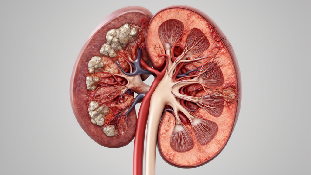
Histopathological evaluation of surgical specimens from 84 nephrectomy procedures revealed the extensive morphological substrate responsible for organ failure. Hematoxylin and eosin staining demonstrated characteristic confluent or scattered granulomatous lesions and extensive caseous necrosis. Cortical sections showed widespread glomerulosclerosis accompanied by vitreous degeneration, lymphocytic infiltration, cystic dilation of Bowman’s space, and severe atrophy of glomerular capillary tufts. The renal tubules exhibited thickened, hyalinized basement membranes and sloughing of necrotic epithelial cells into the tubular lumina, producing structural distortion. Interstitial compartments displayed marked collagenous fibrosis and chronic lymphocytic infiltration. Blood vessels exhibited prominent medial thickening, hyalinization, and lumen narrowing, establishing the histological basis for reduced intrarenal perfusion. In resected ureteral segments, chronic inflammatory cell infiltration, mural fibrosis, luminal stenosis, and occasional granulomas were observed.
Ultrastructural analysis conducted via transmission electron microscopy on resected renal specimens from three nephrectomy patients revealed severe sub-microscopic lesions that explain functional collapse in kidneys lacking gross parenchymal loss. Glomerular capillaries exhibited irregular thickening of their basement membranes alongside extensive fusion or complete effacement of podocyte foot processes. Vascular endothelial cells and mesangial cells displayed marked mitochondrial swelling, endoplasmic reticulum dilation, and an abnormal abundance of lysosomes. Renal tubular structures demonstrated pronounced basement membrane thickening, epithelial detachment, and pronounced mitochondrial injury characterized by balloon-like swelling, localized rupture of cristae, and dissolution of inner and outer mitochondrial membrane boundaries. While the authors emphasized that such ultrastructural damage is not pathognomically specific to mycobacteria—given that chronic ischemia, backpressure, and nonspecific inflammation induce comparable cellular injuries—these alterations confirm that cellular transport and filtration mechanisms break down at an ultrastructural level well before macroscopic parenchymal destruction is recognizable on imaging.
Statistical evaluation examining the relationship between pathological severity and functional loss showed that morphological damage does not cleanly mirror measured functional decline. Spearman’s rank correlation analysis between semi-quantitative histological injury scores and split glomerular filtration rates across 83 patients showed a weak positive association that failed to attain statistical significance (rs equals 0.201, 95 percent confidence interval minus 0.012 to 0.405, p equals 0.068). The authors highlighted significant study limitations, noting an inherent surgical selection bias: because 76.36 percent of participants underwent nephrectomy for end-stage nonfunctional kidneys, the cohort was heavily skewed toward advanced, complicated pathology, preventing generalization to mild or moderate renal tuberculosis managed conservatively with medication. Furthermore, the small number of bilateral cases limited statistical power regarding systemic azotemia, and retrospective data collection precluded multivariable regression adjusting for confounding variables such as patient age, systemic hypertension, diabetes mellitus, or the nephrotoxic effects of antimicrobial agents like aminoglycosides and rifampicin. The authors concluded that preserving renal function in renal tuberculosis demands vigilant functional surveillance for silent contralateral obstruction alongside regimens capable of clearing persistent intrarenal bacilli.
Subject of Research: Clinical and pathological mechanisms of progressive renal function impairment in renal tuberculosis
Article Title: Clinical analysis of factors contributing to renal function impairment in renal tuberculosis
Article References: Li, C., Zhu, G., Liu, J., Zhang, Y., Wang, Q., & Li, W. (2026). Clinical analysis of factors contributing to renal function impairment in renal tuberculosis. Frontiers in Medicine, 13, Article 1932844. https://doi.org/10.3389/fmed.2026.1932844
Image Credits: AI Generated
DOI: 10.3389/fmed.2026.1932844
Keywords: renal tuberculosis, Mycobacterium tuberculosis, glomerular filtration rate, renal dynamic scintigraphy, hydronephrosis, ureteral stricture, silent obstruction, serum creatinine, technetium-99m DTPA, computed tomography urography, glomerulosclerosis, tubular necrosis, transmission electron microscopy, nephrectomy, extrapulmonary tuberculosis, interstitial fibrosis
News Source: Jerry Hayes. (November 21, 2026). Renal Tuberculosis Impairs Kidney Function Through Parenchymal Damage and Silent Obstruction. Scienmag.