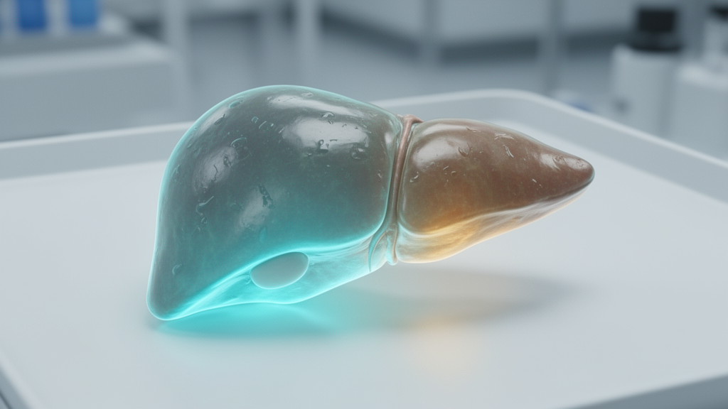
Researchers in Japan have developed an advanced magnetic resonance imaging technique that visualizes the real-time activity of an essential liver enzyme in living organisms for the first time, establishing a novel experimental pathway to detect and track metabolic fatty liver disease. The multidisciplinary investigation, conducted jointly by the National Institutes for Quantum Science and Technology alongside industrial collaborators from Nippon Sanso Corporation, introduced a hyperpolarized carbon-13 molecular probe engineered to track biochemical transformations within liver tissue. Published on November 12, 2026, in the peer-reviewed scientific journal Sensors and Diagnostics, the experimental investigation demonstrated that monitoring paraoxonase enzyme function can reveal critical physiological declines as hepatic steatosis advances. By shifting the diagnostic focus from static anatomical alterations to dynamic enzymatic performance, the collaborative research team demonstrated that specialized imaging can capture subtle functional shifts in mouse models of metabolic dysfunction-associated steatotic liver disease and its severe inflammatory manifestation, metabolic dysfunction-associated steatohepatitis.
Metabolic dysfunction-associated steatotic liver disease is a common condition in which excess fat builds up in the liver in association with metabolic disorders. The condition is estimated to affect about one in three adults. The condition can progress with few obvious warning signs. In some people, the disease progresses to metabolic dysfunction-associated steatohepatitis, liver fibrosis, cirrhosis and even liver cancer.
At the biochemical core of hepatic lipid maintenance is paraoxonase, an enzyme synthesized primarily within liver tissue that plays an indispensable physiological role in regulating lipid metabolism and mitigating damaging oxidative stress. Because of its direct mechanistic participation in mitigating oxidative damage and governing lipid turnover, the operational activity of paraoxonase has emerged as a promising functional biomarker for identifying metabolic perturbations in the liver. Nonetheless, conventional diagnostic methods that rely on standard blood samples face fundamental limitations when attempting to evaluate the true functional capacity of this enzyme. Circulating blood tests cannot accurately reflect how paraoxonase behaves within the internal liver parenchyma, where its enzymatic rate is deeply governed by the complex local biological environment and cellular surroundings.
To overcome the diagnostic blind spots of standard assays, the research group synthesized a specialized metabolic probe utilizing carbon-13-labelled mevalonolactone, formally designated as [1-¹³C]mevalonolactone, tailored specifically to target the enzymatic pathways of paraoxonase. Paraoxonase converts mevalonolactone into mevalonic acid. By measuring the magnetic resonance signals of both the unmetabolized precursor probe and the newly synthesized downstream mevalonic acid product simultaneously, the investigators established a direct comparative framework. Evaluating the ratio and comparative intensities of these two distinct molecular signatures allows researchers to quantitatively visualize and map paraoxonase catalytic activity throughout the liver parenchyma.
The researchers used a technique called hyperpolarization to temporarily boost the probe's MRI signal. Employing a specialized SpinAligner hyperpolarizer apparatus stationed at the Institute for Quantum Life Science within the National Institutes for Quantum Science and Technology, the research group achieved an extraordinary signal enhancement of approximately 230,000-fold over standard magnetic resonance conditions. The hyperpolarized state persisted long enough to reliably trace both the administered mevalonolactone probe and its enzymatic metabolic derivative in living animal subjects.
In living mice, the investigators established that a magnetic resonance scan lasting approximately twenty seconds provided sufficient acquisition time to visualize and quantify hepatic paraoxonase activity comprehensively.
The scientific team rigorously validated their imaging methodology by administering the hyperpolarized molecular probe to laboratory mice subjected to a specialized dietary regimen designed to induce metabolic dysfunction-associated steatotic liver disease and its severe progression to steatohepatitis. When contrasted against healthy control animals maintained on standard nutritional diets, the disease-model mice exhibited a pronounced and progressive reduction in hepatic paraoxonase activity that closely tracked the worsening severity of their liver condition. As steatotic accumulation and inflammatory changes intensified within the hepatic tissue, the corresponding conversion of mevalonolactone to mevalonic acid steadily diminished, producing an unambiguous decline in the recorded metabolic imaging signal. These findings show that the probe can noninvasively detect disease-associated changes in enzyme activity in the living liver with high sensitivity.
Reflecting on the experimental outcomes, Dr. Yoichi Takakusagi, Group Leader at the Institute for Quantum Life Science at the National Institutes for Quantum Science and Technology, emphasized the fundamental diagnostic philosophy driving the research. Takakusagi noted that the research team prioritized creating an imaging methodology capable of revealing what metabolic enzymes are actively doing inside the living body rather than relying solely on blood tests. He explained that their foremost engineering hurdle involved constructing a molecular probe that could generate an exceptionally robust magnetic resonance signal while remaining strictly selective in its response to paraoxonase catalysis. The new probe overcame that hurdle, allowing the researchers to visualize changes in enzyme activity associated with the progression of fatty liver disease.
The broader implications of this imaging framework extend significantly beyond the immediate characterization of metabolic fatty liver disorders. By adapting the fundamental molecular architecture of this carbon-13 probe design toward other specific metabolic enzymes, researchers anticipate that hyperpolarized magnetic resonance imaging could pioneer an entirely new category of medical diagnostics. This approach could open the way to imaging methods that visualize biochemical activity rather than anatomical changes alone. In the longer term, such techniques could contribute to earlier disease detection, assessment of disease progression, and more individualized treatment planning.
The research also represents a focused technological collaboration between academic investigators at the National Institutes for Quantum Science and Technology and industrial researchers from Nippon Sanso Corporation, whose scientists filed related patent applications while providing critical technical development.
Subject of Research: Hyperpolarized carbon-13 MRI visualization of hepatic paraoxonase activity in fatty liver disease mouse models
Article Title: New MRI approach could reveal liver disease earlier
Article References: New MRI approach could reveal liver disease earlier. Original publication (Bibliographic details unavailable.)
Image Credits: AI Generated
DOI: Not provided
Keywords: Hyperpolarized MRI, Paraoxonase, MASLD, MASH, Mevalonolactone, Mevalonic Acid, Carbon-13, Liver Disease, Molecular Imaging, Sensors & Diagnostics, Enzyme Activity, Oxidative Stress, SpinAligner, Metabolic Dysfunction, Yoichi Takakusagi, In Vivo Imaging
News Source: Drew Townsend. (November 21, 2026). Hyperpolarized MRI Probe Visualizes Declining Liver Enzyme Activity in Mouse Disease Models. Scienmag.