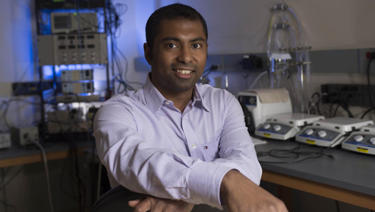
Credit: David Hungate/Virginia Tech
Rengasayee "Sai" Veeraraghavan, a research assistant professor at the Virginia Tech Carilion Research Institute, became the sixth person in the world to receive the George Palade Award from the Microscopy Society of America.
George Palade, a 1974 Nobel Prize Laureate and pioneer in electron microscopy, was credited as the founding father of the contemporary study of cell biology. The award named in his honor recognizes contributions to the field of microscopy and microanalysis in the life sciences by an early career scientist.
"Dr. Veeraraghavan is an extremely gifted biomedical innovator, particularly in the development and application of sophisticated, next-generation imaging tools for the analysis of biological systems, particularly cardiac tissue in health and disease," Michael J. Friedlander, executive director of the Virginia Tech Carilion Research Institute and Virginia Tech's vice president for health sciences and technology. "It is fitting that his contributions are recognized by the prestigious award named after the renowned cell biologist, George Palade, who also revolutionized our ability to visualize and understand cellular structure with unparalleled resolution."
Veeraragahavan studies the structural mechanisms underlying cardiac conduction, in health and disease, at the Virginia Tech Carilion Research Institute's Center for Heart and Regenerative Medicine in the laboratory of Center's director, Rob Gourdie. Veeraragahavan is building upon the work he conducted as a postdoctoral trainee, also at the Virginia Tech Carilion Research Institute, investigating how the physical location of the proteins could potentially support or change conductive behavior between heart cells.
As a postdoctoral trainee, Veeraragahavan began using a new imaging technology called STochastic Optical Reconstruction Microscopy (STORM), which earned the 2014 Nobel Prize in Chemistry, to study proteins in the narrow space between heart cells.
While STORM provided the raw data, Veeraraghavan realized he didn't have a way to process the positions of individual molecules–a necessary step in understanding how the molecules interact. So, he invented a new way to analyze the data.
"With STORM, you don't actually get a picture. You get 3-D positions," Veeraraghavan said. "It's an entirely new type of data, and we had to invent a new method to analyze this information."
Called STochastic Optical Reconstruction Microscopy-based Relative Localization Analysis (STORM-RLA), his novel open-source analysis software allows Veeraraghavan to quickly parse through the locations of single molecules to determine how often and to what degree specific protein clusters interact. It's these interactions that pass the heart's electrical current between cells. When the interactions don't behave as expected, the consequences are deadly.
The visual output from STORM is a graph, providing insight into clusters of proteins. It also accounts for the empty spaces between molecules, resulting in a colossal amount of data that requires several dedicated computers weeks to analyze.
"Previously, the analysis was designed as 'yes or no' in identifying the presence of proteins," Veeraraghavan said, noting that all of the empty spaces would have to be sifted out before researchers could begin questioning how the proteins were interacting. "Now, we're getting a full statistical picture with a lot more useful information."
Veeraragahavan's analysis method asks how far apart the protein clusters are, rather than if a protein is in the imaged space. The resulting data focuses on how, not if, proteins are organized relative to one another.
Beyond investigating how proteins might be regulated to rescue out-of-sync cardiac conduction, Veeraraghavan says the analysis could be used in other fields of study.
"Since the analysis looks at the entire system and identifies all kinds of cluster interactions, it could even work on an astronomical level," Veeraraghavan said. "What works over nanometers works over light years as well."
The George Palade Award recognizes the translational applicability of Veeraraghavan's analysis technique, as well as the potential for future discoveries in several fields of scientific inquiry.
"[He] has used his diverse background in engineering and mathematics to study a complex biological system in the imaging of cardiac junctions," said Christine Brantner, a senior research scientist in electron microscopy at The George Washington University Nanofabrication and Imaging Center, of Veeraraghavan's work. Brantner also co-chairs the awards committee for the Microscopy Society of America. "Sai's publications… have been widely cited and will be of great use to many investigators in the field."
###
The award will be presented on Aug. 7 at the annual Microscopy & Microanalysis meeting, co-sponsored this year by the Microscopy Society of America, the Microanalysis Society, and the International Field Emission Society. The meeting will take place in St. Louis, Missouri.
Media Contact
Ashley WennersHerron
[email protected]
540-526-2002
@vtnews
http://www.vtnews.vt.edu


