Distal left main coronary artery bifurcation evaluation demands a rigorous distinction between physiological lesion significance and downstream procedural complexity, according to an updated narrative review published in Frontiers in Cardiovascular Medicine. Authored by Tong Fangnian, Ma Jun, and Wang Wei from the Department of Cardiology at Wuhan Asian Heart Hospital, the comprehensive synthesis evaluates how conventional two-dimensional coronary angiography frequently misleads interventional cardiologists by failing to depict true plaque distribution, vessel dimensions, and functional hemodynamic resistance. Distal left main disease remains of critical clinical importance because the left main artery supplies an extensive territory of myocardium, meaning revascularization decisions directly alter long-term patient outcomes. To resolve diagnostic discrepancies across modalities, the authors organize existing consensus recommendations and trial evidence into a staged, question-first decision framework spanning untreated native lesions, revascularization selection, percutaneous coronary intervention planning, and the distinct post-procedural assessment of side-branch compromise.
Standard coronary angiography serves as an indispensable anatomical starting point for defining gross bifurcation geometry, vessel tortuosity, and guide catheter engagement, yet its reliance on planar luminograms creates profound clinical blind spots. Planar projection images cannot measure vessel-wall plaque burden, external elastic membrane dimensions, calcification depth, or stent strut apposition, and they frequently misrepresent intermediate stenosis severity due to vessel overlap and radiographic foreshortening. Intravascular ultrasound studies reveal that atherosclerotic plaque extends far beyond what is visible angiographically, clustering predominantly along lateral vessel walls rather than symmetrically encircling the bifurcation carina. Consequently, standard angiographic classifications such as the Medina system fail to reliably capture the true spatial distribution of disease. Furthermore, arterial remodeling introduces substantial discordance between plaque burden and luminal narrowing, as positive outward remodeling preserves lumen cross-sectional area despite marked atheroma accumulation, while negative inward remodeling exaggerates apparent luminal loss. In intermediate left main disease, visual angiographic severity therefore correlates only weakly with physiological pressure measurements and cross-sectional intravascular imaging.
To interrogate the functional consequences of anatomical narrowing, interventional cardiologists employ pressure-derived indices, primarily hyperemic fractional flow reserve and resting instantaneous wave-free ratio. Established clinical cutoffs identify functional ischemia at a fractional flow reserve of 0.80 or less and an instantaneous wave-free ratio of 0.89 or less. However, the authors caution that distal left main lesions cannot be analyzed like isolated single-vessel stenoses because hyperemic flow across the left main bifurcation is heavily influenced by downstream vascular resistance. An abnormal distal wire pressure reflects the cumulative pressure losses incurred across both the left main trunk and the interrogated daughter vessel, meaning a depressed pressure ratio does not locate ischemia exclusively to the left main trunk. Conversely, severe downstream narrowing in either the left anterior descending or left circumflex artery restricts hyperemic blood flow, paradoxically reducing the measured trans-lesion pressure gradient across an upstream left main stenosis and making it appear falsely non-ischemic. Operators must verify pressure sensor equalization, eliminate guide-catheter damping, check for hydrostatic and electrical drift, and perform meticulous hyperemic or resting pullbacks across both branches to separate focal left main pressure loss from diffuse downstream disease.
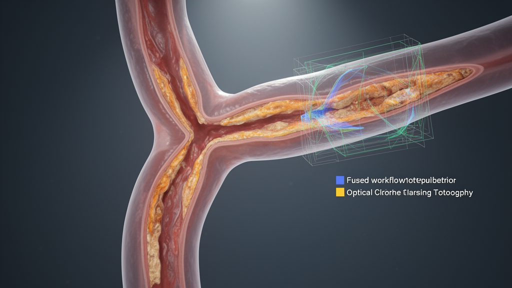
Intravascular imaging modalities, comprising intravascular ultrasound and optical coherence tomography, provide cross-sectional vessel characterization but operate under distinct technical capabilities and physical limitations. Intravascular ultrasound features superior acoustic tissue penetration, rendering it well suited for deep vessel sizing, mapping external elastic membrane diameters, delineating plaque burden, and selecting healthy landing zones in large-caliber left main segments. In the multicenter prospective LITRO study evaluating intermediate left main lesions, a minimal lumen area of 6.0 square millimeters or greater safely supported revascularization deferral. However, the review emphasizes that this threshold is an observational study benchmark rather than a universally validated biological cutoff for ischemia, noting that studies in East Asian cohorts have observed physiological significance at minimal lumen areas near 4.5 square millimeters for isolated ostial or shaft lesions. In contrast, optical coherence tomography provides substantially higher axial resolution, excelling in the microscopic detection of calcified plaque morphology, stent strut apposition, edge dissections, and guidewire recrossing positions. Nevertheless, optical coherence tomography requires active blood clearance and suffers from shallower light penetration, complicating full-depth vessel visualization in dilated left main anatomies.
The review delineates four non-exclusive, overlapping clinical scenarios where diagnostic modalities frequently provide conflicting assessments. In the first scenario, a severe-appearing angiographic stenosis yields non-ischemic physiology, necessitating systematic verification of pressure calibration, downstream branch resistance, and intravascular ultrasound adjudication before deferral is considered. In the second scenario, angiographically mild or intermediate narrowing masks high-risk anatomical features, such as dense calcification or heavy plaque burden identified on intravascular imaging; these features dictate technical lesion preparation and device selection if percutaneous revascularization is indicated, but they do not serve as standalone triggers for stenting. In the third scenario, intermediate angiographic stenosis exhibits abnormal functional ischemia, compelling operators to confirm whether the pressure drop originates in the left main or downstream arteries before choosing between revascularization pathways. In the fourth scenario, physiological discordance emerges between the left anterior descending and left circumflex branches, driven by divergent downstream lesion severity, unequal subtended myocardial mass, or asymmetric plaque distribution across the bifurcation carina.
To navigate these ambiguities, the authors outline an evidence-informed, sequential clinical workflow that prioritizes fundamental clinical questions before procedural execution. Step one mandates defining the overarching clinical setting, explicitly separating the pre-intervention assessment of untreated native left main bifurcations from the post-procedural evaluation of a jailed side branch. Step two determines whether the lesion is functionally or anatomically significant using high-quality angiography, selective branch pullbacks, and adjunctive intravascular ultrasound, while strictly avoiding treating procedural complexity or calcification as surrogates for ischemia. Step three determines whether to defer revascularization under optimized medical therapy or proceed with revascularization based on concordant objective findings. If revascularization is indicated, step four requires selecting between percutaneous coronary intervention and coronary artery bypass grafting by evaluating overall coronary complexity, patient operative risk, and Heart Team consensus prior to committing to specific catheter-based preparation. Step five establishes the specific percutaneous strategy, favoring provisional single-stent implantation for most non-complex anatomies while reserving planned two-stent approaches for selected complex true bifurcations. Finally, step six governs imaging-guided stent optimization, the proximal optimization technique, and post-dilatation.
A central clinical distinction within the review is the absolute separation between untreated native left main bifurcations and the post-intervention assessment of a jailed left circumflex ostium following crossover stenting. When an operator implants a stent across the left main into the left anterior descending artery, carina shift and plaque snowplowing frequently produce marked angiographic pinching of the circumflex ostium. Observational and physiological studies demonstrate that this visual pinching is largely geometric and rarely corresponds to true hemodynamic compromise or diminished coronary flow. The authors emphasize that post-crossover angiographic pinching must never trigger uncritical rescue stenting in stable patients exhibiting preserved flow and absent electrocardiographic changes. Selective side-branch pressure wire interrogation and intravascular imaging should instead verify functional compromise or significant malapposition before additional stents are placed. Furthermore, converting to rescue side-branch stenting after crossover PCI must not be conflated with an upfront, planned two-stent bifurcation technique selected prior to initial stent deployment.
Evaluating the contemporary clinical trial landscape, the authors highlight substantial heterogeneity across landmark randomized studies and caution against pooling disparate trial results into universal clinical mandates. In the randomized EBC MAIN trial of true left main bifurcations, stepwise provisional stenting demonstrated three-year major adverse cardiac event rates of 23.5 percent compared with 29.5 percent for systematic dual stenting, alongside target-lesion revascularization rates of 8.3 percent and 15.6 percent, respectively. Conversely, the DKCRUSH-V trial evaluated a distinct, highly complex true left main population managed with operator-dependent double-kissing crush techniques, reporting three-year target-lesion failure of 8.3 percent with DK crush versus 16.9 percent with provisional stenting. Because these trials enrolled patient cohorts with markedly different bifurcation complexities and anatomical side-branch lesion lengths, their divergent outcomes reflect population-specific treatment responses rather than contradictory evidence. Similarly, while large trials such as OCTOBER and RENOVATE-COMPLEX-PCI demonstrated clinical benefits from intravascular imaging guidance in complex percutaneous interventions, the left-main-specific OPTIMAL trial found no significant reduction in patient-oriented composite events with routine intravascular ultrasound guidance at 2.9 years.
Observational registries providing the evidentiary foundation for revascularization deferral similarly carry structural methodological limitations that preclude causal claims of equivalence. In the prospective LITRO registry of 354 patients, deferring revascularization in 179 of 186 patients with an intravascular ultrasound minimal lumen area of at least 6.0 square millimeters yielded a two-year cardiac death-free survival rate of 97.7 percent, compared with 94.5 percent among revascularized patients. In an earlier prospective cohort of 213 equivocal left main lesions by Hamilos and colleagues, fractional-flow-reserve-directed medical care produced an estimated five-year overall survival of 89.8 percent versus 85.4 percent with coronary artery bypass grafting. The observational iLITRO-EPIC07 registry examined combined fractional flow reserve and instantaneous wave-free ratio assessment across 300 patients, resolving index discordance with intravascular ultrasound and observing a primary composite event rate of 8.3 percent among deferred patients versus 13.3 percent among revascularized patients over 20 months. However, the authors underscore that non-randomized treatment assignment, observational selection bias, and wide statistical confidence intervals mean these findings cannot establish clinical equivalence between medical deferral and revascularization.
Ultimately, the review emphasizes that the proposed multimodality framework serves as an evidence-informed organizational synthesis rather than a prospectively validated diagnostic classification or algorithmic clinical rule. The diagnostic sequence is applicable primarily to stable patients with intermediate or equivocal narrowing and must never delay emergent revascularization in unstable acute coronary syndromes, hemodynamic shock, or life-threatening ongoing ischemia. Technical hurdles such as severe stenosis preventing intravascular imaging catheter passage require careful lesion preparation with micro-balloons only after revascularization has been decided, avoiding pre-dilation for diagnostic imaging alone. The authors conclude that future prospective randomized investigations must directly evaluate whether implementing this integrated, question-first multimodality workflow improves decision-making quality, procedural precision, and long-term clinical endpoints beyond current guideline-directed standard practice.
Subject of Research: Interventional multimodality assessment and revascularization decision workflow for distal left main coronary artery bifurcation lesions
Article Title: Multimodality assessment of LM bifurcations when angiography misleads: an interventional workflow integrating angiography, physiology and intravascular imaging for left main bifurcation lesions
Article References: Fangnian, T., Jun, M., & Wei, W. (2026). Multimodality assessment of LM bifurcations when angiography misleads: an interventional workflow integrating angiography, physiology and intravascular imaging for left main bifurcation lesions. Frontiers in Cardiovascular Medicine, 13, 1994428. https://doi.org/10.3389/fcvm.2026.1994428
Image Credits: AI Generated
DOI: 10.3389/fcvm.2026.1994428
Keywords: left main coronary artery, bifurcation lesions, coronary angiography, fractional flow reserve, instantaneous wave-free ratio, intravascular ultrasound, optical coherence tomography, percutaneous coronary intervention, coronary artery bypass grafting, provisional stenting, DK crush, hemodynamic ischemia, plaque morphology, minimal lumen area, proximal optimization technique, jailed side branch, cardiovascular intervention
News Source: Ophelia Keating. (November 20, 2026). Review Outlines Multimodality Workflow for Complex Left Main Coronary Bifurcation Assessment. Scienmag.