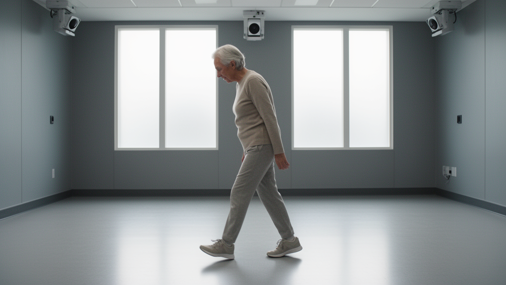
In adults with degenerative spinal disease, worsening sagittal balance during walking stems from a forward shift in posture rather than a breakdown in trunk and pelvic coordination, according to a cross-sectional instrumented gait analysis published in the Journal of Orthopaedic Surgery and Research. The optoelectronic motion investigation revealed that while static standing radiographs often conceal postural adaptations by compressing compensated patients into the same tier as balanced individuals, dynamic walking kinematics uncover a distinct forward-shifted operating point while preserving the underlying trunk–pelvis coupling.
Evaluating sagittal imbalance has traditionally relied on standing full-spine radiographs, which capture static postural alignment but fail to record how the trunk and pelvis coordinate dynamically during self-paced movement. The research team sought to determine whether instrumented gait analysis could distinguish between different radiographic balance and compensation categories where static measures typically collapse into two broad tiers. Specifically, the authors examined whether progressive spinal imbalance represents a mechanical breakdown in coordination between the trunk and pelvis or instead reflects an intact coupling pattern functioning around an anteriorly shifted posture.
The cross-sectional investigation evaluated forty-eight adults diagnosed with degenerative spinal disease. Each participant underwent both standing full-spine radiography and three-dimensional optoelectronic gait analysis while walking at a self-selected pace. To categorize spinal balance, the researchers employed two radiographic classification axes based on sagittal vertical axis decompensation, defined as a sagittal vertical axis exceeding 50 millimeters, and activated pelvic compensation, marked by a pelvic tilt greater than 20 degrees. From this framework, three distinct patient groups were analyzed: a balanced cohort without compensation comprising fifteen individuals, a balanced cohort with activated compensation consisting of eighteen individuals, and a decompensated cohort with insufficient compensation containing twelve individuals.
Biomechanical assessments utilized marker-based tracking to measure trunk inclination, denoted as TL, alongside pelvic tilt, denoted as PT, across both static standing and dynamic walking conditions. To evaluate within-pass trunk–pelvis coordination, the investigators calculated the Spearman correlation between trunk inclination and pelvic tilt, where negative correlation values represented an antiphase coordination pattern. This dynamic relationship was benchmarked against a rigid-trunk geometric null model. Diagnostic accuracy and threshold discrimination were evaluated through receiver operating characteristic curve analysis using the Benjamini–Hochberg procedure to control for multiplicity, with correlation strengths interpreted against established published scales.
Kinematic tracking during walking demonstrated a progressive, monotonic increase in trunk inclination across the three clinical tiers. Mean walking trunk inclination measured 7.7 degrees in balanced uncompensated patients, rose to 10.3 degrees in balanced compensated patients, and reached 16.1 degrees in decompensated patients, demonstrating a statistically significant progression with a p-value below 0.001. In sharp contrast, static standing trunk inclination separated only the most severe group, failing to distinguish between the two balanced tiers.
Despite the progressive forward lean observed during walking, the fundamental coordination between the trunk and pelvis remained intact across the patient cohort. Antiphase trunk–pelvis coupling was documented in all forty-five classified participants, significantly exceeding the rigid-trunk geometric null model. Furthermore, this antiphase relationship showed no statistically significant difference across the three clinical groups, yielding a p-value of 0.261. These observations confirm that advancing sagittal imbalance reflects an anterior translation of the operating point within an intact motor pattern rather than a mechanical dissolution of trunk–pelvis coupling.
Static evaluations also provided predictive utility for identifying severe misalignment, as each additional degree of standing trunk inclination was associated with a 1.49-fold increase in the odds of radiographic decompensation, with a 95 percent confidence interval spanning 1.17 to 1.89. Receiver operating characteristic analysis identified an optimal standing trunk inclination threshold of approximately 5.8 degrees, which achieved an area under the curve of 0.904, accompanied by a sensitivity of 0.92 and a specificity of 0.73. Notably, nurse-assessed gait instability correlated significantly with standing sagittal vertical axis and standing trunk inclination, with q-values of 0.008 and 0.038 respectively, but showed no correlation with dynamic gait parameters. The authors concluded that while brief standing measures can triage decompensation, instrumented gait analysis serves as an effective functional stress test that reveals hidden dynamic balance adaptations in degenerative spinal disease.
Subject of Research: Instrumented gait analysis of trunk–pelvis coupling and sagittal balance in degenerative spinal disease
Article Title: Walking reveals a forward-shifted trunk–pelvis operating point that standing alignment conceals: trunk–pelvis coupling is preserved across radiographic balance–compensation groups in degenerative spinal disease — a cross-sectional instrumented gait analysis
Article References: Li, X., Fan, Z., Pan, F., Zhang, H., Wang, S., Wang, D., Wang, Y., Kong, C., Li, J., Chen, X., & Lu, S. (2026). Walking reveals a forward-shifted trunk–pelvis operating point that standing alignment conceals: trunk–pelvis coupling is preserved across radiographic balance–compensation groups in degenerative spinal disease — a cross-sectional instrumented gait analysis. Journal of Orthopaedic Surgery and Research. https://doi.org/10.1186/s13018-026-07340-y
Image Credits: AI Generated
DOI: 10.1186/s13018-026-07340-y
Keywords: degenerative spinal disease, instrumented gait analysis, trunk inclination, pelvic tilt, trunk–pelvis coupling, sagittal balance, sagittal imbalance, standing radiographs, optoelectronic motion analysis, sagittal vertical axis, pelvic compensation, decompensation, gait kinematics, antiphase coupling, gait instability, receiver operating characteristic, spine biomechanics
News Source: Ophelia Keating. (November 20, 2026). Walking Reveals Concealed Forward Trunk Shift in Degenerative Spinal Disease. Scienmag.