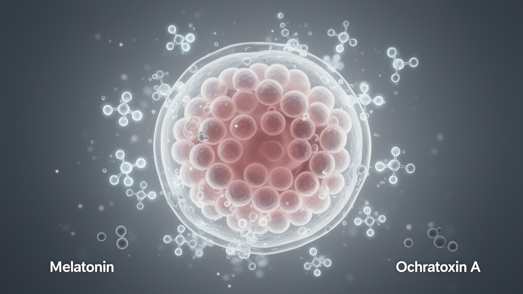
Exposure to the food-borne mycotoxin ochratoxin A impairs preimplantation porcine embryo development, while subsequent melatonin supplementation mitigates this damage, according to laboratory findings published in Cell Biology and Toxicology. The study evaluated how toxic exposure disrupts developmental competence following in vitro fertilization and explored whether melatonin could alleviate the resulting cellular stress.
Ochratoxin A is a contaminant known to compromise female reproductive function, yet the underlying biological pathways driving its toxicity during early embryonic stages have remained unclear. Researchers focused specifically on endoplasmic reticulum stress and quality-control mechanisms to understand the disruption occurring within the first 48 hours after in vitro fertilization.
To test these effects, researchers exposed developing porcine embryos to ochratoxin A concentrations of 0.5, 1.0, or 5.0 micromolar during their initial 48-hour developmental window. Following the exposure interval, the embryos were cultured either with or without melatonin supplementation until reaching the blastocyst stage, while tunicamycin served as a positive control for endoplasmic reticulum stress.
Initial evaluations demonstrated that ochratoxin A exposure directly hindered embryonic cleavage before any melatonin was introduced. As development proceeded, the mycotoxin significantly suppressed blastocyst formation, reduced overall embryo quality, and triggered elevated levels of programmed cell death, or apoptosis.
Transcriptomic profiling via RNA sequencing revealed that ochratoxin A exposure enriched genes associated with oxidative stress, apoptosis, autophagy, and the unfolded protein response. Molecular assessments confirmed an increased abundance of GRP78 alongside elevated phosphorylation of PERK and eIF2alpha, accompanied by the accumulation and nuclear localization of ATF5.
The toxin also compromised endoplasmic reticulum architecture and maintenance, causing reduced SEC62 abundance and weaker ER-Tracker staining intensity. While signals for the autophagy marker LC3B increased in exposed embryos, their spatial overlap with endoplasmic reticulum structures declined, reflecting disrupted coupling necessary for ER-phagy.
Subsequent supplementation with melatonin notably counteracted these cellular defects during post-exposure culture. Embryos provided with melatonin showed improved blastocyst development and overall quality, reduced apoptosis, and attenuated signaling across the PERK, eIF2alpha, and ATF5 stress pathway.
Melatonin treatment also partially restored SEC62 abundance and helped re-establish structural organization related to ER-phagy in the treated embryos. The findings demonstrate that post-exposure melatonin intervention can alleviate ochratoxin A-induced developmental toxicity while altering key markers of endoplasmic reticulum stress and quality control in porcine embryos.
Methodologically, the researchers separated evaluation stages to distinguish immediate toxic impacts from later developmental consequences. Cleavage was assessed specifically before introducing melatonin, whereas blastocyst formation, overall embryo quality, apoptosis, transcriptomic changes, endoplasmic reticulum stress signaling, and organelle quality control markers were evaluated during subsequent embryonic development.
These collective observations indicate that ochratoxin A exposure reduces porcine preimplantation development while provoking distinct changes across PERK, eIF2alpha, and ATF5 signaling alongside SEC62 associated endoplasmic reticulum quality control markers. Ultimately, administering melatonin following toxic exposure improved developmental outcomes in the porcine embryos, an improvement that was consistently associated with measurable modifications in these critical stress and autophagy related markers.
Subject of Research: Melatonin alleviation of ochratoxin A embryotoxicity in porcine embryos
Article Title: Melatonin alleviates ochratoxin A-induced toxicity and restores developmental competence of porcine embryos, possibly via PERK signaling
Article References: Ha, H.-W., Koo, D.-B., & Park, H.-J. (2026). Melatonin alleviates ochratoxin A-induced toxicity and restores developmental competence of porcine embryos, possibly via PERK signaling. Cell Biology and Toxicology. https://doi.org/10.1007/s10565-026-10364-z
Image Credits: AI Generated
DOI: 10.1007/s10565-026-10364-z
Keywords: ochratoxin A, melatonin, porcine embryos, in vitro fertilization, blastocyst development, endoplasmic reticulum stress, PERK signaling, eIF2alpha, ATF5, GRP78, SEC62, ER-phagy, apoptosis, unfolded protein response, mycotoxin toxicity, embryo quality
News Source: Drew Townsend. (November 20, 2026). Melatonin Alleviates Ochratoxin A Toxicity in Developing Porcine Embryos. Scienmag.