A decellularized bovine pericardial bioscaffold clinically employed in cardiovascular surgery provides a permissive, non-directive microenvironment that sustains human mesenchymal stromal cell adhesion, proliferation, and multipotency without forcing premature lineage commitment, according to an in vitro investigation published in the Journal of Materials Science: Materials in Medicine. Tissue engineering strategies across musculoskeletal reconstruction, cardiac repair, and broader regenerative medicine rely heavily on biomaterial platforms capable of supporting transplanted progenitor cells during critical periods of post-implantation attachment and tissue integration. However, a persistent challenge in biomaterial translation has been ensuring that the biological substrate does not inadvertently trigger unscheduled differentiation in seeded cells or host progenitors. By combining functional cellular assays with high-throughput global transcriptomic profiling, researchers demonstrated that human stromal cells cultured directly on the engineered collagen matrix retain their characteristic regenerative secretome and broad stem cell identity while altering the expression of merely fifty-seven genes out of more than thirteen thousand mapped transcripts.
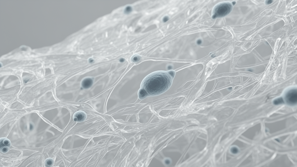
The biomaterial examined in the study is CardioCel, an acellular, collagen-rich matrix derived from bovine pericardium that was originally developed for congenital and adult cardiovascular procedures, including cardiac valve reconstruction, septal defect closures, and complex ventricular tunnel repairs. The scaffold is manufactured utilizing proprietary ADAPT technology, an engineered process that integrates antigen-depleting decellularization, controlled low-toxicity glutaraldehyde chemical crosslinking, and a specialized anti-calcification treatment formulated to reduce cytotoxicity while ensuring long-term mechanical durability under hemodynamic stress. Anatomically, the processed bioscaffold preserves the native tri-layered architecture of mammalian pericardium, comprising a rough and fibrous external surface, an organized middle parietal layer, and a smooth visceral lining that natively faces the myocardium. This structural retention provides distinct surface topographies and fibrillar orientations, providing a physiologically relevant foundation to evaluate how primary human reparative cells physically engage and adapt to clinical biomaterial surfaces.
To investigate the cellular response across distinct progenitor lineages, the research team evaluated primary human adipose-derived mesenchymal stromal cells and human bone marrow-derived mesenchymal stromal cells obtained from commercial donors aged twenty to forty-five years. Both cell populations conformed to established surface marker criteria, exhibiting confirmed expression of standard mesenchymal markers including CD29, CD44, CD73, CD90, CD105, and CD166, while testing entirely negative for hematopoietic and endothelial lineage markers CD14, CD31, CD34, and CD45. Prior to scaffold evaluation, working cell stocks expanded through passages five to eight were validated on conventional tissue culture plastic under controlled serum conditions, where they consistently formed confluent spindle-shaped monolayers with doubling times below twenty-five hours and maintained baseline viability above seventy percent. In parallel baseline testing, working cell stocks verified their standard capacity to undergo adipogenic, chondrogenic, and osteogenic differentiation under lineage-specific chemical induction.
High-resolution scanning electron microscopy and confocal immunofluorescence imaging revealed that human mesenchymal stromal cells actively adapted their morphology across the distinct topographies of the decellularized pericardial scaffold within two days of seeding. On the textured fibrous surface, seeded cells formed direct physical contacts with individual collagen bundles, extending elongated cellular processes that bridged microscopic pores and conformed to the undulating matrix architecture. In contrast, stromal cells seeded on the smoother visceral surface organized into uniform, flattened monolayers reflecting the planar underlying matrix. Confocal microscopy of cell nuclei and filamentous actin cytoskeletal networks stained with phalloidin confirmed that cells distributed at varying three-dimensional depths within the porous fibrous matrix, whereas visceral-seeded cells remained strictly aligned within a single horizontal plane, proving that primary human stromal cells can establish robust mechanical and structural attachment across the diverse physical microenvironments present within the scaffold.
Quantitative metabolic adherence and proliferation assays demonstrated that the bioscaffold rapidly supports viable cellular accumulation in a density-dependent manner. Cell adherence evaluations conducted at two and seven hours using colorimetric tetrazolium bioreduction demonstrated statistically significant attachment across all experimental densities compared with acellular scaffold blanks. Within two hours of initial seeding, low-density cultures exhibited optical density increases exceeding fifteen percent, whereas high-density seeding produced absorbance increases up to 56.1 percent in adipose-derived cells and 64.1 percent in bone marrow-derived populations. Continued monitoring of metabolic activity over twenty-four, forty-eight, and seventy-two hours revealed significant, progressive cellular accumulation across all density groups, with highest-density cultures displaying metabolic signal increases of 75.7 percent and 87.3 percent for adipose and bone marrow stromal cells, respectively. Direct optical cell counting across time points was precluded by the inherent autofluorescence of the glutaraldehyde-crosslinked matrix, but complementary nuclear imaging and metabolic kinetics established that seeding densities of at least 13,000 cells per square centimeter ensure rapid, stable engraftment without requiring protracted pre-incubation.
Crucially for regenerative applications, cells maintained on the pericardial matrix preserved their full trilineage differentiation potential when exposed to specialized biochemical induction cocktails over extended two- to three-week culture periods. Following fourteen days of adipogenic culture, Oil Red O histological staining revealed abundant cytoplasmic neutral lipid droplets within scaffold-attached cells, comparable to plastic controls. Chondrogenic microtissues cultured in chondrogenic medium formed rounded cartilage-like nodules synthesizing abundant glycosaminoglycan-rich extracellular matrix that stained intensely with Alcian Blue dye, with bone marrow-derived stromal cells exhibiting visibly greater nodule formation than adipose-derived lines. Under osteogenic cues, von Kossa staining identified dark mineralized calcium phosphate salt deposits distributed across the scaffold surfaces after twenty-four days, confirming functional osteoblastic maturation. Importantly, uninduced control cultures maintained on the bioscaffold in baseline growth medium exhibited minimal spontaneous lineage transformation, demonstrating that the structural matrix remains permissive to directed differentiation signals without imposing unsolicited fate instructions.
To evaluate whether contact with the crosslinked pericardium altered progenitor identity at the molecular level, researchers performed global stranded messenger RNA sequencing on Illumina platforms, profiling cells maintained on the scaffold against paired plastic cultures after forty-eight hours of growth. Following read alignment to the human GrCh38 reference genome and multi-factor differential expression modeling with DESeq2 to statistically isolate substrate effects from intrinsic donor-source variation, the investigators observed remarkable transcriptomic stability. Out of 13,810 mapped transcripts, only fifty-seven genes met the rigorous threshold for statistically significant differential expression, defined by an adjusted probability value below 0.05 and a log2 fold change of at least two. Principal component analysis and sample-to-sample distance heatmaps confirmed that the primary source of transcriptomic variance was biological cell origin, while the scaffold substrate introduced only secondary, modest transcriptional adjustments that clustered tightly among biological replicates.
Among the fifty-seven altered transcripts, twenty-two genes were significantly upregulated, representing an active adaptive program focused primarily on cell-matrix adhesion, membrane transport, and microenvironmental sensing. Gene ontology enrichment analysis highlighted significant clusters localized to the plasma membrane and extracellular region, featuring biological adhesion and cellular responses to endogenous organic stimuli and altered oxygen levels. Other upregulated genes included angiopoietin-like 4 (ANGPTL4), an endothelial survival factor and matrix binder induced 2.4-fold, solute carrier family 16 member 6 (SLC16A6, induced 4.3-fold), carbonic anhydrase 9 (CA9, 3.7-fold), gap junction protein beta 2 (GJB2, 2.9-fold), bone morphogenetic protein 2 (BMP2, 2.5-fold), and developmental Wnt signaling regulators NOTUM, NKD2, and WNT7B.
Conversely, thirty-five transcripts showed significant downregulation, pointing to an adaptive dampening of de novo matrix synthesis, pro-fibrotic signaling, and inflammatory pathways. The down-regulation of endogenous structural and adhesive transcripts, including beta-1,3-galactosyltransferase 2 (B3GALT2, down four-fold), hyaluronan and proteoglycan link protein 1 (HAPLN1, down 3.6-fold), and cadherin 4 (CDH4, down 2.2-fold), suggests that stromal cells reduce energetic expenditure on matrix synthesis when provided with an abundant, pre-existing collagenous framework. Furthermore, reductions in pappalysin 2 (PAPPA2, down 2.5-fold), a protease that cleaves IGFBP-5, and connective tissue growth factor (CTGF, down 2.1-fold) were accompanied by significant repression of pro-inflammatory interleukin 26 (IL26, down 2.3-fold) and annexin A3 (ANXA3, down 2.3-fold), a calcium-binding regulator whose repression aligns with the engineered anti-calcification properties of the scaffold. Crucially, the broad regenerative secretome of mesenchymal stromal cells remained robustly preserved, with key trophic and angiogenic regulators, including VEGFA, TGFA, TGFB1, TGFB2, IL1B, IL6, ANGPT1, THBS2, PDGFA, HBEGF, and matrix metalloproteinases MMP1, MMP2, MMP3, MMP9, and MMP13, maintaining stable expression across both scaffold and plastic conditions.
The study authors emphasize that these molecular and phenotypic findings establish decellularized bovine pericardium as a highly biocompatible cell delivery substrate, while also cautioning that clinical translation requires careful matching of scaffold biomechanics to target anatomical sites. Nonetheless, the findings establish that in vitro mesenchymal stromal cell assays serve as a sensitive predictive screening benchmark to verify that decellularized surgical biomaterials support viable progenitor retention without compromising stemness.
Subject of Research: Evaluation of decellularized bovine pericardium bioscaffold compatibility and transcriptomic stability in human mesenchymal stromal cells
Article Title: Adaptation of mesenchymal stromal cells to culture on collagen-based bioscaffold
Article References: Ong, H. T., Vargas-Landin, D. B., Redmond, S. L., Neethling, L., Rhodes, D. I., & Dilley, R. J. (2026). Adaptation of mesenchymal stromal cells to culture on collagen-based bioscaffold. Journal of Materials Science: Materials in Medicine, 37(1), Article 104. https://doi.org/10.1007/s10856-026-07069-4
Image Credits: AI Generated
DOI: 10.1007/s10856-026-07069-4
Keywords: mesenchymal stromal cells, CardioCel, decellularized bioscaffold, bovine pericardium, transcriptome profiling, multipotency, trilineage differentiation, ADAPT technology, regenerative medicine, cell adhesion, extracellular matrix, RNA sequencing, ITGA2, tissue engineering, cell delivery platform, anti-calcification
News Source: Drew Townsend. (November 20, 2026). Decellularized Pericardium Bioscaffold Preserves Human Stem Cell Function Without Directing Fate. Scienmag.