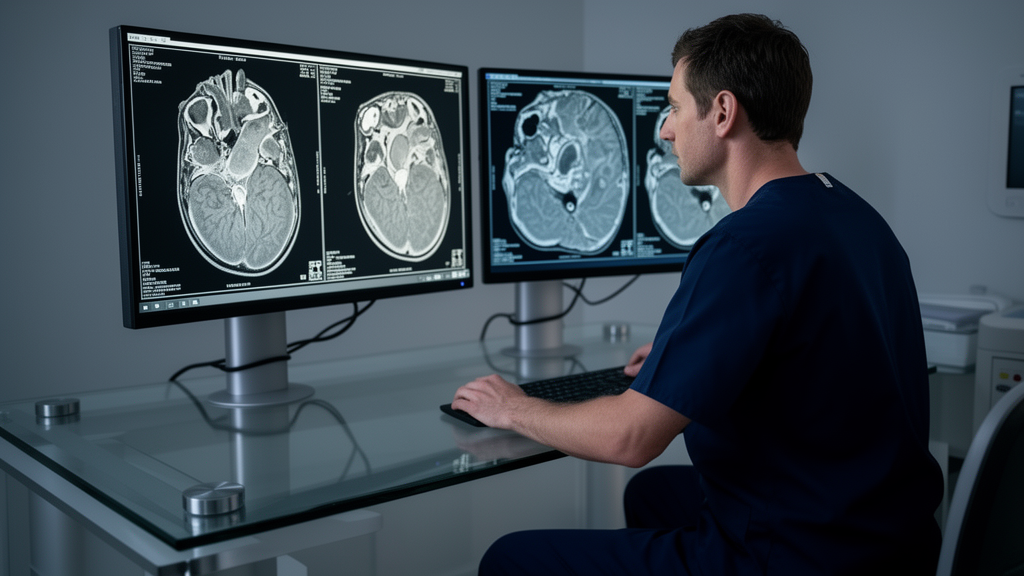
A retrospective study of noncontrast cardiac computed tomography scans demonstrates that specialized three-dimensional deep learning architectures can segment the left atrial appendage and quantify its volume with high spatial fidelity and minimal measurement bias. Published in the Journal of Cardiovascular Imaging, the investigation examined imaging datasets from 452 patients aged 60 years or older who had undergone evaluation for unstable angina at a diagnostic imaging center in northern Paraná state, Brazil. By evaluating four distinct U-Net variants against manual annotations performed by an expert cardiologist, the researchers determined that deep learning models can perform complex anatomical delineations directly from scans that do not require intravenous iodinated contrast media. The authors highlighted that while the left atrial appendage represents a primary anatomical site of intracardiac thrombus formation, routine noncontrast scans have historically underutilized this structure due to high anatomical variability and the absence of standardized, automated clinical post-processing tools.
The clinical motivation for developing automated segmentation models on noncontrast cardiac computed tomography stems from the structural and physiological significance of the left atrial appendage in cardiovascular pathology. The appendage is a complex, tubular structure that experiences pathological dilation in settings involving endothelial injury, a proinflammatory status, and blood stasis, which collectively elevate the risk of thromboembolic events and atrial fibrillation. While noncontrast computed tomography is routinely acquired in clinical environments to evaluate coronary artery calcium scoring, detailed structural quantification of the appendage on these scans has remained technically difficult. Standard noncontrast protocols utilize anisotropic voxel dimensions and relatively thick image slices, which limits multiplanar reconstruction and obscures the ostial boundary between the appendage and the left atrial chamber in oblique and sagittal planes. Consequently, prior volumetric studies have predominantly relied on contrast-enhanced coronary computed tomography angiography, creating an operational barrier for patients presenting with contraindications to contrast agents or severe renal compromise.
To evaluate whether deep learning could resolve these challenges, the investigators retrospectively collected institutional data spanning from August 2019 to November 2023 under an institutional ethics protocol aligned with the Checklist for Artificial Intelligence in Medical Imaging guidelines. The final analyzed cohort comprised 452 patients who had undergone both noncontrast cardiac computed tomography and contrast-enhanced angiography as part of a standardized chest pain diagnostic pathway. The cohort presented a median age of 68 years with an interquartile range of 63 to 73 years, and 282 patients, or 62.4 percent, were male. The median body mass index across the group was 28 kilograms per square meter, with an interquartile range of 26 to 32 kilograms per square meter. Within the cohort, the most prevalent recorded comorbidities included systemic hypertension in 80.3 percent of participants, dyslipidemia in 40.7 percent, and type 2 diabetes mellitus in 36.1 percent, representing an older clinical population prone to structural atrial remodeling.
The patient inclusion protocol enforced strict physiological and clinical parameters, requiring all individuals to be at least 60 years of age with a verified clinical diagnosis of unstable angina, an estimated creatinine clearance of at least 30 milliliters per minute per 1.73 square meters, a resting heart rate of no more than 70 beats per minute during acquisition, absence of significant physical agitation, and high overall image quality. The specific age cutoff of 60 years was selected based on established epidemiological data, including findings from the Framingham Heart Study demonstrating that atrial fibrillation incidence approximately doubles with each successive decade past age 60. This epidemiological trajectory is closely linked with progressive structural remodeling of the atria, including tissue dilation and morphofunctional alterations that accelerate from the sixth decade onward. Setting this inclusion threshold aligned with standardized clinical risk stratification frameworks such as the CHA2DS2-VASc score while mitigating developmental anatomical heterogeneity found across younger demographics.
All noncontrast computed tomography scans were generated using an 80-detector Aquilion Lightning scanner from Canon Medical Systems, operating with prospective reconstruction and electrocardiographic monitoring synchronized to 75 percent of the cardiac cycle, corresponding to mid-to-late ventricular diastole. The acquisition parameters included a tube voltage of 120 kilovolts, a tube current of 300 milliamperes, an image matrix of 512 by 512, an FC12 convolution kernel reconstruction filter, and a slice thickness of 3.0 millimeters. Paired contrast-enhanced scans were captured using retrospective gating with radiation dose modulation between 40 and 80 percent, reconstructed at 80 percent of the cardiac cycle with a 0.5-millimeter slice thickness and a 0.3-millimeter interslice gap. The authors acknowledged that because the left atrial appendage attains its true maximal volume during end-systole, image acquisition synchronized to mid-diastole introduced a minor systematic underestimation of maximal chamber dimensions across all examined subjects.
Establishing the reference ground truth required rigorous manual segmentation conducted by a cardiologist with more than a decade of specialized experience in clinical cardiology, cardiac computed tomography, and cardiac magnetic resonance imaging. The expert manual annotations were delineated using Horos version 3.3.6 and 3D Slicer version 5.0.3 according to international expert recommendations for planning transcatheter left atrial appendage occlusion. The manual segmentation protocol explicitly excluded the body of the left atrium at the ostial margin, as well as the pulmonary veins and neighboring mediastinal structures. Because anisotropic acquisition precluded reliable visualization of the ostium in sagittal views, the boundary was standardized based on axial transition zones where the appendage visibly narrows from the main atrial cavity. To avoid bias, the cardiologist remained entirely blinded to all automated algorithmic predictions and volumetric measurements generated throughout the investigation.
The deep learning pipeline implemented four volumetric convolutional neural network architectures built upon the U-Net encoder-decoder paradigm: standard UNet3D, Residual-UNet3D, 3D Attention-UNet, and Res16-PAC-UNet. Each model featured a contracting encoder path with four downsampling stages driven by 2 by 2 by 2 max pooling, followed by an expansive decoder path with three upsampling stages using transposed convolutions with a stride of 2, culminating in a 1 by 1 by 1 convolution to produce final binary segmentation maps. The base UNet3D applied repeated 3 by 3 by 3 convolutions with batch normalization, rectified linear unit activation, and a dropout rate of 0.2. The Residual-UNet3D integrated skip residual connections to facilitate gradient backpropagation. The 3D Attention-UNet incorporated spatial and channel attention gates into skip connections to emphasize salient regions of interest, while the Res16-PAC-UNet employed multiscale pyramid atrous convolutions with dilation rates of 6, 12, and 18 to expand receptive fields.
Image preprocessing standardized the noncontrast volumetric scans by resampling the CT volumes to a uniform matrix of 128 by 128 by 64 voxels via Z-axis spline interpolation, balancing spatial feature retention with hardware limits on a single graphics processing unit. Voxel intensities were clipped and normalized to a soft-tissue Hounsfield unit window ranging from 50 to 350, purposely isolating structural tissue while excluding hyperdense calcifications or thrombi. Training leveraged fivefold patient-level cross-validation across the 452 scans to prevent data leakage between training and validation folds. In each fold, networks were trained for 100 epochs using the Adam optimizer with a fixed learning rate of 0.001 and soft Dice loss as the objective function, supplemented by data augmentation including small three-dimensional rotations between negative and positive 1 degree, brightness and contrast shifts within 20 percent, and grid distortions up to 0.3.
Spatial segmentation accuracy across the validation folds yielded mean Dice similarity coefficients of 78.44 plus or minus 1.93 for UNet3D, 78.97 plus or minus 0.79 for Residual-UNet3D, 79.07 plus or minus 1.43 for 3D Attention-UNet, and 77.68 plus or minus 1.47 for Res16-PAC-UNet. A Friedman test across the cross-validation folds demonstrated no statistically significant difference in segmentation performance among the four architectures, returning a chi-square value of 7.80 with a p-value of 0.0503. Subsequent post hoc Wilcoxon signed-rank tests adjusted with Bonferroni correction yielded adjusted p-values of 0.375 or higher across all pairwise comparisons. In volumetric analysis calculated from original DICOM voxel dimensions, all models showed strong, statistically significant linear correlations with expert manual volumes (p less than 0.001), led by the 3D Attention-UNet with a Pearson correlation coefficient of 0.800, while the Residual-UNet3D yielded a coefficient of 0.762.
Complementary Bland–Altman agreement analysis revealed minimal volumetric bias across all evaluated architectures, demonstrating mean differences of negative 0.24 milliliters for UNet3D, positive 0.14 milliliters for Residual-UNet3D, negative 0.17 milliliters for 3D Attention-UNet, and negative 0.18 milliliters for Res16-PAC-UNet. From an operational perspective, automated inference required an average execution time of approximately 2 seconds per scan on a standard workstation equipped with an AMD Ryzen 3900X central processing unit and a single GeForce RTX 3090 Ti graphics processing unit. This throughput reduced the total analytical duration for the entire 452-patient cohort to under 20 minutes, compared to an estimated 75 to 150 hours required for manual tracing at standard clinical rates of 10 to 20 minutes per patient. In a preliminary validation cohort of 57 patients, comparing noncontrast estimates against contrast-enhanced computed tomography angiography revealed a minimal mean volumetric difference of 0.14 milliliters.
The authors detailed several methodological constraints inherent to the study design, emphasizing that the single-center retrospective format and reliance on scanners from a single vendor with a uniform acquisition protocol may limit generalizability across broader clinical settings. Furthermore, because image acquisition occurred at 75 percent of the cardiac cycle, measurements reflect mid-diastole rather than true end-systolic maximal chamber dimensions. The pipeline also relied exclusively on internal cross-validation without an independent external test cohort, which may modestly overstate performance metrics. Finally, the study was designed strictly for anatomical volume quantification rather than intraluminal thrombus detection and was restricted entirely to patients aged 60 and older with unstable angina, warranting future multicenter investigations to confirm broader diagnostic validity.
Subject of Research: Deep learning segmentation and volume quantification of the left atrial appendage using noncontrast cardiac computed tomography
Article Title: Deep learning models for segmentation and quantification of left atrial appendage volume using noncontrast cardiac computed tomography
Article References: Santos, D. A. M., de Oliveira Teixeira, L., Massago, M., da Alvarez Silva, S., Jr., Gurgel, S. J. T., Rochitte, C. E., da Costa, Y. M. E. G., & de Andrade, L. (2025). Deep learning models for segmentation and quantification of left atrial appendage volume using noncontrast cardiac computed tomography. Journal of Cardiovascular Imaging, 33(1), Article 16. https://doi.org/10.1186/s44348-025-00058-1
Image Credits: AI Generated
DOI: 10.1186/s44348-025-00058-1
Keywords: Left Atrial Appendage, Noncontrast Cardiac CT, Deep Learning, 3D U-Net, Medical Image Segmentation, Cardiac Imaging, Volume Quantification, 3D Attention-UNet, Residual-UNet3D, Dice Similarity Coefficient, Bland-Altman Analysis, Atrial Fibrillation, Thrombus Risk, Computer-Aided Diagnosis, Cardiovascular Imaging, Aquilion Lightning
News Source: Blake Davidson. (November 19, 2026). Deep Learning Models Accurately Measure Left Atrial Appendage Volume Without Contrast Agents. Scienmag.