A prospective clinical study investigating visceral adiposity around the heart has revealed that epicardial fat volume measured using artificial intelligence on cardiac computed tomography is independently linked to markers of left ventricular diastolic dysfunction, but not to other conventional indicators of systemic target organ damage. The investigation, conducted in individuals diagnosed with nonobstructive coronary artery disease, evaluated whether indexed epicardial fat volume correlates with impairment across multiple organ domains including renal filtration, vascular stiffness, and cardiac mechanical relaxation. The research team identified that greater accumulation of epicardial adipose tissue relates specifically to deterioration in early diastolic tissue velocities and elevated left ventricular filling pressures, while showing no statistically significant associations with kidney function markers or peripheral arterial stiffening.
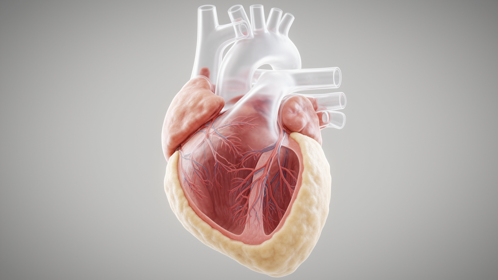
Epicardial adipose tissue covers roughly eighty percent of the cardiac surface and functions as an active visceral organ capable of synthesizing and secreting a variety of bioactive molecules, hormones, and cytokines that exert direct local effects on cardiovascular tissues. Because epicardial fat is situated directly adjacent to the myocardium without intervening fascial layers, molecular cross-talk occurs unhindered through paracrine and vasocrine pathways. While previous research has linked epicardial adiposity to metabolic syndrome, waist circumference, and the anatomical severity of coronary atherosclerosis, prior investigations evaluating its relationship with wider target organ damage produced variable results, largely due to reliance on two-dimensional ultrasound thickness measurements rather than comprehensive three-dimensional volumetric assessment.
To provide standardized quantification of this fat depot, investigators enrolled a prospective cohort of consecutive patients undergoing electrocardiogram-gated coronary computed tomography angiography and transthoracic echocardiography at Seoul Metropolitan Government Seoul National University Boramae Medical Center between February 2017 and March 2021. From an initial screening pool of 372 consecutive individuals, the research protocol excluded patients who presented with an interval exceeding thirty days between imaging and laboratory tests, active chest pain during examination, a left ventricular ejection fraction below fifty percent, significant valvular disease, or obstructive coronary artery disease defined by fifty percent or greater luminal stenosis. Following the systematic application of these rigorous criteria, 75 eligible patients with nonobstructive coronary artery disease were retained for full cross-sectional evaluation.
The study cohort presented with a mean participant age of 57.1 years and included 39 male patients, representing 52.0 percent of the sample. Participants exhibited an average body mass index of 26.1 kilograms per square meter, a mean brachial systolic blood pressure of 123.5 millimeters of mercury, and a mean diastolic blood pressure of 73.2 millimeters of mercury. Comorbid conditions identified across the population included dyslipidemia in 22.7 percent, hypertension in 21.3 percent, and diabetes mellitus in 13.3 percent of cases. Concomitant pharmacotherapies documented among participants encompassed statin therapy in 30.7 percent, calcium channel blockers in 16.0 percent, renin-angiotensin system blockers in 16.0 percent, and beta-blockers in 13.3 percent, reflecting standard medical management in this patient group.
Measurement of epicardial fat volume was executed from retrospectively electrocardiogram-gated computed tomography datasets acquired on a 128-slice scanner, with all examinations completed within an average interval of 5.6 days from echocardiographic and laboratory evaluations. The researchers segmented adipose tissue located between the myocardium and the visceral pericardium, bounded superiorly by the pulmonary trunk bifurcation and extending inferiorly to the pericardial base. Automated three-dimensional segmentation was performed using deep learning software employing 3D U-Net neural network architecture, identifying voxels exhibiting attenuation values between minus 190 and minus 30 Hounsfield units. Two experienced cardiothoracic radiologists subsequently conducted minimal boundary refinement to eliminate internal voids and ensure anatomical consistency before standardizing absolute volume against body surface area derived via the Mosteller formula.
The cohort displayed an average unadjusted epicardial fat volume of 155.6 cubic centimeters, yielding a body surface area-adjusted index of 91.4 cubic centimeters per square meter. Concurrently, comprehensive transthoracic echocardiography documented preserved systolic performance, with a mean left ventricular ejection fraction of 60.1 percent and an average left ventricular end-diastolic dimension of 50.0 millimeters. Key parameters of diastolic filling and relaxation demonstrated a mean deceleration time of early mitral inflow of 200.0 milliseconds, an average ratio of early to late peak mitral inflow velocity of 0.95, a mean septal early diastolic tissue velocity of 6.5 centimeters per second, and a mean ratio of early mitral inflow to annular tissue velocity of 10.5.
Beyond cardiac imaging, the clinical protocol gathered parallel metrics across other target organ systems, observing a mean estimated glomerular filtration rate of 100.6 milliliters per minute per 1.73 square meters, a mean hemoglobin A1c of 6.2 percent, and a mean total cholesterol level of 181.0 milligrams per deciliter. Peripheral arterial elasticity and vascular resistance evaluations recorded an average brachial-ankle pulse wave velocity of 1430 centimeters per second alongside an average ankle-brachial index of 1.15. Albumin-to-creatinine ratios were determined from random spot urine samples, and tricuspid regurgitation velocities were evaluated via continuous-wave Doppler to provide a complete systemic assessment of target organ damage.
Statistical evaluation began with linear correlation analyses, which demonstrated that indexed epicardial fat volume increased alongside patient chronological age, presenting a moderate positive correlation coefficient of 0.567. When investigators conducted partial correlation analyses controlling specifically for the confounding influence of age, indexed epicardial fat volume maintained a significant negative correlation with septal early diastolic tissue velocity (r = -0.489) and a significant positive correlation with the ratio of early mitral inflow to annular velocity (r = 0.256). In stark contrast, no significant correlations emerged between indexed fat volume and estimated glomerular filtration rate, urine albumin-to-creatinine ratio, brachial-ankle pulse wave velocity, ankle-brachial index, or tricuspid regurgitation velocity after controlling for age.
To verify whether the relationships observed with left ventricular diastolic parameters persisted independently of established cardiometabolic risks, the investigators constructed multivariable linear regression models adjusting for age, body mass index, hypertension, diabetes mellitus, smoking status, and renal filtration capacity. Within these fully adjusted models, indexed epicardial fat volume remained an independent predictor of septal early diastolic tissue velocity, with a regression coefficient of minus 0.0003, as well as an independent predictor of the ratio of early inflow to annular velocity, yielding a coefficient of 0.0606. These findings confirmed that local adipose accumulation around the heart retains a distinct association with impaired ventricular relaxation and elevated filling pressures irrespective of systemic hemodynamic or metabolic covariates.
Diagnostic accuracy assessments using receiver operating characteristic curve analysis further highlighted the capability of computed tomography-derived indexed fat volume to discriminate clinically defined thresholds of diastolic dysfunction. For detecting a reduced septal early diastolic tissue velocity below 7 centimeters per second, indexed fat volume achieved an area under the curve of 0.750, with an optimal cut-off value of 95.78 cubic centimeters per square meter providing 88.2 percent sensitivity and 56.8 percent specificity. For detecting an elevated ratio of early inflow to annular tissue velocity greater than 12, the metric yielded an area under the curve of 0.692, where an optimal cut-off threshold of 91.68 cubic centimeters per square meter demonstrated 71.4 percent sensitivity and 64.8 percent specificity.
The biological mechanisms accounting for this localized connection involve both molecular and mechanical factors inherent to the epicardial fat depot. Because the tissue shares an unobstructed microcirculation with myocardial layers, adipokines and pro-inflammatory signaling proteins such as tumor necrosis factor alpha, interleukin 6, and monocyte chemoattractant protein 1 can diffuse directly into adjacent myocardium, fostering microvascular inflammation and myocardial fibrosis. Furthermore, excessive anatomical volume within the unyielding pericardial sac may impose mechanical constraint against diastolic filling, while myocardial infiltration of free fatty acids from surrounding fat stores can trigger localized lipotoxicity and stiffen ventricular muscle.
The authors noted several important constraints regarding the interpretation and generalizability of their results. Conducted at a single medical center with a final sample size of 75 patients, the study possessed limited statistical power to rule out weaker associations with non-cardiac target organ parameters, and unmeasured confounding factors cannot be completely excluded despite multivariable statistical adjustments. Because the cross-sectional design captured data at a single time point without prospective follow-up, the study design precludes drawing causal inferences or determining long-term clinical prognosis. Nevertheless, the findings confirm that volumetric epicardial fat quantified through deep learning algorithms serves as a specific anatomical biomarker preferentially linked to cardiac diastolic performance in nonobstructive coronary disease.
Subject of Research: Association of indexed epicardial fat volume with left ventricular diastolic function in nonobstructive coronary artery disease
Article Title: Association of deep learning–derived epicardial fat volume with target organ damage in subjects with nonobstructive coronary artery disease
Article References: Kim, M. Y., Kim, H.-L., Chun, E. J., Choi, Y. R., & Jin, K. N. (2025). Association of deep learning–derived epicardial fat volume with target organ damage in subjects with nonobstructive coronary artery disease. Journal of Cardiovascular Imaging, 33(1), Article 17. https://doi.org/10.1186/s44348-025-00062-5
Image Credits: AI Generated
DOI: 10.1186/s44348-025-00062-5
Keywords: epicardial fat volume, epicardial adipose tissue, cardiac computed tomography, deep learning segmentation, nonobstructive coronary artery disease, left ventricular diastolic dysfunction, septal e-prime velocity, E over e-prime ratio, target organ damage, arterial stiffness, brachial-ankle pulse wave velocity, estimated glomerular filtration rate, body surface area, receiver operating characteristic, coronary computed tomography angiography, echocardiography, tissue doppler imaging
News Source: Blake Davidson. (November 19, 2026). Deep Learning Epicardial Fat Volume Associates Specifically With Cardiac Diastolic Dysfunction. Scienmag.