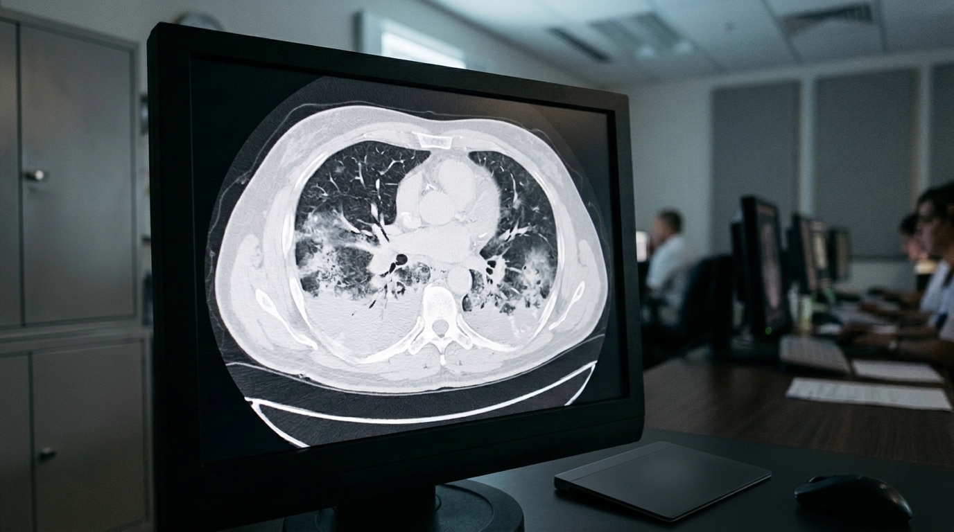
A comprehensive scientific review published in Intensive Care Medicine Experimental details how advanced X-ray computed tomography enables the quantitative assessment of regional lung mechanical properties in acute respiratory distress syndrome, or ARDS. ARDS is an acute and often lethal form of hypoxemic respiratory failure marked by severe inflammation, noncardiogenic pulmonary edema, and marked structural heterogeneity across lung tissue. Standard clinical management relies heavily on endotracheal intubation and supportive mechanical ventilation to maintain gas exchange. However, conventional bedside monitoring assesses respiratory function through global measurements, including airway pressure, flow waveforms, respiratory system compliance, driving pressure, and mechanical power. Because ARDS inflicts patchy, uneven damage throughout the pulmonary parenchyma, global metrics often fail to capture the severe localized strains and stresses that cause ventilator-induced lung injury, known as VILI.
The review emphasizes that the spatial heterogeneity inherent to ARDS breaks the conventional clinical assumption that globally delivered mechanical ventilation exerts uniform physical forces throughout the injured lung. Even in a healthy respiratory system, tissue compliance, opening pressures, and mechanical time constants vary naturally because of nonlinear alveolar pressure-volume relationships and vertical intrapleural pressure gradients. In ARDS, this variation becomes dramatically amplified by dependent fluid accumulation, gravity, and localized inflammatory exudate. Classical pulmonary physiology established by Mead and colleagues demonstrated that neighboring alveoli behave interdependently rather than as isolated functional balloons, sharing physical forces across shared interalveolar septa. When injured, collapsed, or fluid-filled alveoli sit adjacent to aerated lung units, alveolar interdependence amplifies local distending stresses well beyond global transpulmonary pressures, creating focal stress concentrations that perpetuate parenchymal tearing and cyclic injury.
Quantitative X-ray computed tomography has emerged as a central modality to characterize these structural derangements across whole lung volumes with high spatial resolution. Modern volumetric CT scanners capture whole-organ datasets in sub-second acquisition intervals, generating calibrated voxel-based maps of attenuation expressed in Hounsfield units, where air measures minus 1000 Hounsfield units and pure water registers at 0 Hounsfield units. In human clinical CT scanners, a typical reconstruction voxel measuring approximately one cubic millimeter encompasses roughly 160 alveoli. By applying linear attenuation equations, researchers compute the precise volume fraction of gas and tissue within each individual voxel. This enables researchers and clinicians to map regional aeration distributions across the entire lung, pinpointing localized areas of overdistension, functional aeration, or complete collapse.
Segmentation of the lung boundaries from mediastinal and chest wall structures serves as the primary gateway for regional quantitative analysis. While healthy lungs display sharp contrast differences that make threshold-based automated segmentation straightforward, ARDS presents substantial segmentation challenges because dense areas of atelectasis, consolidation, and edema blur the boundaries between parenchyma and neighboring chest structures. To overcome these limitations, modern automated pipelines increasingly rely on deep learning architectures, specifically convolutional neural networks, to delineate injured pulmonary borders rapidly and reliably. Once segmented, parenchyma can be systematically classified into four distinct functional compartments originally standardized by Gattinoni: hyperaerated tissue below minus 901 Hounsfield units, normally aerated tissue between minus 900 and minus 501 Hounsfield units, poorly aerated tissue between minus 500 and minus 101 Hounsfield units, and non-aerated tissue above minus 100 Hounsfield units.
Density-based CT analyses of ARDS have fundamentally transformed intensive care concepts by establishing the paradigm of the baby lung, describing the sharply reduced volume of normally aerated lung tissue that remains functional and receives the bulk of delivered tidal ventilation. This aerated compartment typically occupies non-dependent ventral zones, whereas dependent dorsal zones demonstrate dense, non-aerated consolidation. Furthermore, volumetric CT imaging performed at varying static airway pressures differentiates the baby lung from recruitable lung tissue. Recruitable lung represents non-aerated regions that can be reopened through increased positive end-expiratory pressure or recruitment maneuvers. Studies demonstrate enormous interpatient variability in recruitability: some patients exhibit substantial reaeration of collapsed dependent tissue when airway pressures are raised from 5 to 45 centimeters of water, while other patients show virtually no recruitment despite having similar average densities, explaining why standardized ventilatory protocols fail to yield uniform patient outcomes.
To evaluate dynamic tissue movement rather than static density alone, investigators utilize deformable image registration, an advanced computational technique that establishes voxel-to-voxel spatial correspondence across images acquired at different inflation pressures, volumes, or respiratory phases. Unlike rigid registration, which accounts only for translation and rotational shifts, deformable registration models the complex, non-rigid, and non-uniform distortion of lung tissue. The resulting vector displacement field describes the local trajectory of every volume element between the reference and moving images. Registration accuracy is rigorously evaluated using expert-defined anatomical landmarks, inverse-consistency checks, and standardized reference datasets, ensuring that the computed spatial transformations accurately represent true anatomical tissue deformation across healthy and severely diseased lung states.
From these deformable vector fields, researchers calculate the local Jacobian determinant, a scalar metric representing the local volumetric expansion ratio between two physiological states. A Jacobian determinant greater than 1 indicates regional volumetric expansion, a value less than 1 reflects local compression, and values near 1 indicate static tissue volume. Dynamic respiratory-gated four-dimensional CT combined with deformable registration demonstrates that following acute lung injury, regional strain distributions become markedly heterogeneous. Regions of elevated strain frequently concentrate within aerated tissue borders directly adjacent to collapsed, non-yielding units. Furthermore, by coupling regional volume differentials with localized estimates of superimposed pleural pressure derived from esophageal balloon manometry, researchers can calculate regional respiratory compliance on a voxel-by-voxel basis across the pulmonary volume.
Beyond standard Hounsfield density thresholds, advanced texture analysis methods characterize complex parenchymal architecture in ARDS. Supervised machine-learning algorithms, such as the adaptive multiple feature method, classify lung parenchyma into detailed texture classes including normal tissue, ground-glass opacities, reticular patterns, bronchovascular bundles, and solid consolidation. Complementary mathematical approaches, including quadtree decomposition algorithms, compute the mean homogeneous volume of compliance, specific ventilation, and parenchymal strain fields, providing objective numerical metrics of structural and functional heterogeneity. However, calculating voxel-level strain in severely injured lungs introduces complex physiological considerations; voxels representing non-inflated or poorly aerated alveoli at functional residual capacity possess very small initial volume denominators, which can produce artificially inflated mathematical strain values unless corrected through accurate recruitment estimates.
The integration of quantitative CT datasets with computational biomechanical modeling offers powerful avenues to infer physical variables that cannot be directly measured in living patients. In a representative modeling pipeline, three-dimensional central airway trees extracted from segmented clinical CT scans serve as geometric boundary conditions for algorithmic branching networks that generate peripheral airways down to terminal acini. Regional CT measurements of aeration, tissue density, and recruitability parameterize localized resistance, elastance, and impedance within forward finite element or lumped-parameter simulations. These CT-informed computational models calculate the regional distribution of tidal airflow, gas exchange, and intraparenchymal stress concentrations under simulated mechanical ventilation settings, enabling mechanistic hypothesis testing and simulated evaluations of patient-specific ventilatory strategies.
Despite its profound analytical power, quantitative CT imaging faces practical obstacles that constrain its routine adoption for daily ventilator titration in critical care units. Transporting critically ill patients from the intensive care unit to a radiology suite carries substantial risk, with complications and adverse events documented in up to 70 percent of transfers. Additionally, standard thoracic CT delivers an ionizing radiation dose of approximately 7 millisieverts, limiting the safety of repeated longitudinal acquisitions, while intravenous contrast agents pose risks of hypersensitivity reactions and acute nephrotoxicity. Deformable registration and high-resolution computational fluid simulations also demand extensive computational resources that currently prevent real-time bedside execution.
Promising technological innovations are actively addressing these translational barriers. Emerging photon-counting detector CT systems measure the individual energy levels of incoming X-ray photons directly, eliminating electronic noise, enhancing spatial resolution, improving soft-tissue contrast, and enabling multi-energy spectral characterization at significantly reduced radiation doses. Concurrently, mobile cone-beam CT scanners are being adapted for direct bedside deployment in intensive care units, circumventing the physiological hazards of intrahospital transport. Although current portable scanners show lower signal-to-noise ratios, elevated scatter, and less reliable Hounsfield calibration than standard multidetector helical systems, ongoing advancements in reconstruction algorithms and detector sensitivity continue to narrow this performance gap.
In summary, quantitative X-ray computed tomography bridges the divide between global clinical mechanics and the microscopic cellular forces that drive ventilator-induced lung injury. While clinical trials have yet to demonstrate a definitive survival advantage for CT-guided ventilatory adjustments over standardized low-tidal-volume ventilation, CT imaging remains the scientific reference standard for characterizing the physical state of the injured lung. By combining refined segmentation, deformable image registration, texture classification, and patient-specific computational modeling, quantitative CT provides essential mechanistic insights into ARDS pathophysiology, establishing a robust framework for developing safer, individualized mechanical ventilation paradigms.
Subject of Research: Quantitative computed tomography assessment of regional lung mechanics in acute respiratory distress syndrome
Article Title: Quantitative assessment of lung mechanical properties in ARDS using X-ray computed tomography
Article References: Gao, J., Garberi, R., Akor, E. A., Perchiazzi, G., & Kaczka, D. W. (2026). Quantitative assessment of lung mechanical properties in ARDS using X-ray computed tomography. Intensive Care Medicine Experimental, 14(1), Article 99. https://doi.org/10.1186/s40635-026-00946-w
Image Credits: AI Generated
DOI: 10.1186/s40635-026-00946-w
Keywords: acute respiratory distress syndrome, computed tomography, ventilator-induced lung injury, mechanical ventilation, lung mechanics, deformable image registration, baby lung, recruitability, Jacobian determinant, regional strain, texture analysis, biomechanical modeling, Hounsfield units, photon-counting CT, pulmonary aeration
News Source: Ophelia Keating. (November 19, 2026). Computed Tomography Quantifies Regional Lung Mechanics and Injury Patterns in ARDS. Scienmag.