Biomechanical researchers have demonstrated that artificial neural networks can predict compressive knee contact force time series during overground walking using joint kinematics extracted from a single standard video camera combined with four demographic variables. In a newly published investigation evaluating the markerless framework against laboratory-grade optical motion capture and musculoskeletal simulations, investigators found that predicted total, medial, and patellofemoral forces correlated strongly with reference calculations across the stance phase of walking. However, the system exhibited substantial inaccuracy when predicting lateral knee contact forces and displayed large variations in error across individual subjects. These performance discrepancies demonstrate that while camera-based joint loading analysis represents a technically viable alternative to specialized laboratory infrastructure, the current implementation remains insufficiently reliable for individual-level clinical assessment, diagnostic decision-making, or guided rehabilitation monitoring without substantial methodological refinements.
Understanding the internal physical loading experienced by the knee joint during daily locomotion is essential because excessive or abnormal contact forces contribute directly to the progressive structural degeneration of articular cartilage. Walking represents an ubiquitous, highly repetitive activity that imparts cyclical contact stresses across both the tibiofemoral and patellofemoral compartments of the knee joint. Under clinical conditions, measuring these internal contact forces experimentally has historically required surgically implanted instrumented knee prostheses, making direct measurement completely unviable for healthy populations or individuals undergoing conservative joint therapy. Consequently, researchers rely on noninvasive musculoskeletal modeling and simulation to calculate compressive joint contact forces. Conventional simulations require individuals to perform walking trials within specialized motion capture laboratories equipped with multi-camera optoelectronic tracking arrays and embedded ground reaction force plates. The prohibitive financial expense, lengthy anatomical marker attachment protocols, and specialized technical expertise needed to operate these motion facilities severely restrict access, preventing routine assessment of knee loading in outpatient clinics, physiotherapy centers, and athletic environments.
To overcome the operational bottlenecks of motion laboratories, biomechanists have turned toward lightweight markerless motion capture solutions driven by computer vision and deep learning architectures. Free markerless methods leverage human pose estimation algorithms that detect two-dimensional anatomical keypoints from ordinary digital imagery and infer three-dimensional coordinates through multi-camera triangulation or single-camera depth lifting techniques. Concurrently, machine learning models have demonstrated that time-series contact forces can be predicted directly from kinematics or ground reaction forces, bypassing computationally demanding static optimization routines. However, video cameras cannot record external ground reaction forces, which are traditionally deemed essential alongside kinematic trajectories to estimate musculoskeletal joint contact forces accurately. While prior investigations explored whether artificial neural networks could estimate peak loading values from reduced kinematic inputs, the new study evaluated whether continuous, stance-phase knee contact force waveforms could be predicted directly from three-dimensional joint angles derived from monocular, single-camera recordings when informed by basic demographic metrics.
To train the predictive neural network architecture, the research team aggregated five established motion capture datasets encompassing 208 healthy participants and 5,009 individual overground walking stance phases. This training cohort included 119 male and 89 female subjects aged between 19 and 84 years, with body mass index measurements ranging between 17 and 41. Raw marker trajectories and ground reaction force plate measurements had been processed through an integrated OpenSim and MATLAB musculoskeletal pipeline utilizing the Rajagopal body model equipped with the Lerner knee model. For every walking stance phase, defined by a vertical ground reaction force threshold of 50 Newtons, inverse kinematics and static optimization were executed to extract reference contact forces and joint kinematics. The resulting reference data yielded sixteen distinct kinematic time series across both the stance limb and contralateral swinging leg, covering lumbar, pelvis, hip, knee, and ankle rotations, alongside subject body mass, height, age, and biological sex.
The predictive architecture chosen through five-fold cross-validation consisted of one-dimensional convolutional neural networks employing a convolutional kernel size of five. Rather than directly outputting final joint forces in a single step, each trained network incorporated two dedicated functional components: a convolutional block that extracted preliminary force trajectories from the sixteen joint angle time series, and a demographic mask block that scaled the preliminary force curve based on participant body mass, height, age, and sex. Dedicated models were trained independently for four compressive force components: total tibiofemoral force, medial tibiofemoral force, lateral tibiofemoral force, and patellofemoral contact force. Model weights were optimized using eighty percent of the aggregated motion data, representing 170 participants and 4,006 individual walking trials, while the remaining twenty percent was reserved for early stopping criteria to prevent overfitting during training.
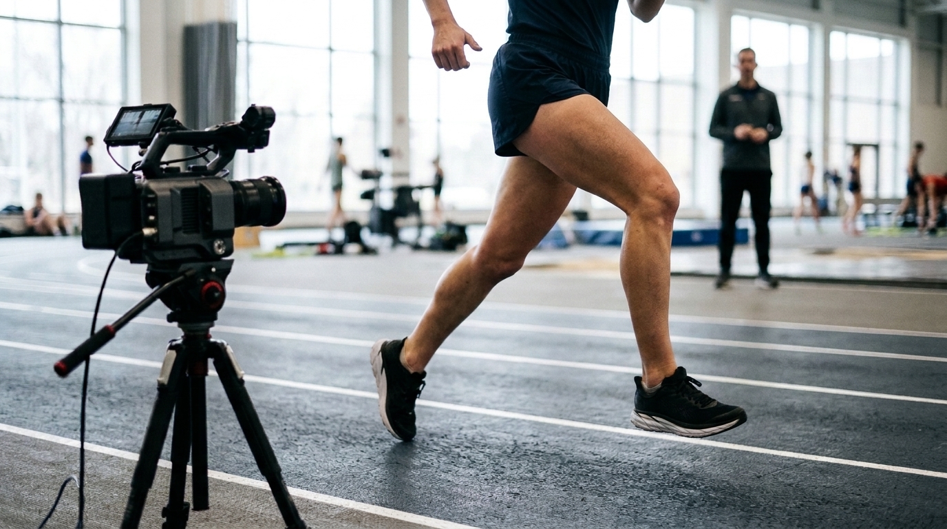
Generating markerless kinematic inputs from single-camera recordings required adapting the Portable Biomechanics Laboratory workflow, which utilizes the MeTRAbs human pose estimation model to extract 87 full-body three-dimensional keypoint trajectories directly from video frames. To translate keypoints into anatomically constrained joint rotations, the investigators fitted a modified Rajagopal skeletal model within the MuJoCo physics engine using a bilevel numerical optimization loop executed for 30,000 iterations per trial. Dependent knee coordinates were explicitly removed from the physics model to prevent unconstrained coordinate optimization relative to knee flexion. The resulting markerless joint angles were smoothed using a zero-phase low-pass digital filter at a 12-Hertz cutoff frequency. These processed kinematic curves and participant demographic metrics were then fed into the trained convolutional networks to predict stance-phase markerless knee contact force time series without requiring any force plate measurements.
The entire workflow was validated using the independent BioCV test dataset, which contains simultaneous high-speed monocular video, fifteen-camera optical motion capture, and synchronized force plate recordings from twelve healthy young adults aged 19 to 38 years. Across sixty analyzed walking trials recorded from a three-quarter waist-height camera perspective, the predicted markerless force curves yielded root mean square errors of 47 percent of body weight for total tibiofemoral force, 40 percent for medial tibiofemoral force, 30 percent for lateral tibiofemoral force, and 29 percent for patellofemoral force. Pearson linear correlation coefficients between predicted and reference curves exceeded 0.80 for total, medial, and patellofemoral contact forces, demonstrating strong profile tracking throughout the stance phase. Nevertheless, lateral tibiofemoral force predictions correlated only moderately at R = 0.64 and exhibited negative coefficients of determination across multiple participants.
A critical technical failure identified in the workflow involved the severe underestimation of the initial lateral tibiofemoral force peak during the first half of the stance phase. Because the mean coefficient of determination for lateral contact forces dropped below zero, the convolutional network performed worse than a basic naive statistical model that merely outputs the mean time series value. The authors observed that reference lateral forces within the training dataset exhibited exceptionally high natural variability during early stance. This wide distribution likely encouraged the optimization algorithm to default toward predicting average values rather than learning precise individual kinematic mappings. Furthermore, systematic differences existed between the training datasets and the BioCV test cohort; first loading peaks occurred earlier and with lower magnitudes in the training data, inducing an early temporal bias across all predicted peaks in the markerless evaluations.
When comparing their motion-capture-driven reference predictions against prior machine learning studies, the authors highlighted distinct methodological trade-offs. Previous neural network models published by other research groups achieved lower normalized errors by utilizing simpler feedforward designs, training on smaller cohorts, or incorporating anatomical frontal-plane tibiofemoral alignment angles as explicit network inputs. However, direct numerical comparisons between published models remain difficult due to conflicting error normalization conventions, differences in walking speeds, varied musculoskeletal modeling assumptions, and the frequent absence of openly available codebases. In this study, prediction errors increased noticeably when switching from optical motion capture kinematics to video-derived markerless kinematics, confirming that errors originating during single-camera pose estimation and physics-based model fitting propagate directly into the downstream kinetic neural network.
Several operational limitations restrict the clinical translation of this single-camera framework. Kinematic estimation proved computationally demanding, requiring approximately four hours on a modern high-performance laptop to process five short walking videos through 30,000 optimization iterations, contrasted with only one second required for neural network force inference. Furthermore, the validation cohort was restricted to young, healthy individuals walking under controlled, well-illuminated laboratory conditions wearing close-fitting attire, meaning findings cannot be generalized to older adults, clinical cohorts, or individuals with obesity whose soft tissue movement hampers computer-vision keypoint tracking. Most critically, because the study evaluated only absolute loading accuracy during normal walking, it did not demonstrate whether the workflow possesses sufficient sensitivity to detect within-person loading alterations following targeted gait retraining, leaving its ultimate clinical utility unproven until validated in larger, more diverse patient populations.
Subject of Research: Prediction of knee joint contact forces from monocular video and demographic information
Article Title: Predicting Knee Contact Forces During Walking From Monocular Video and Demographic Information
Article References: Lavikainen, J., Kim, H., Pagnon, D., Arokoski, J., & Stenroth, L. (2026). Predicting Knee Contact Forces During Walking From Monocular Video and Demographic Information. Annals of Biomedical Engineering. https://doi.org/10.1007/s10439-026-04461-7
Image Credits: AI Generated
DOI: 10.1007/s10439-026-04461-7
Keywords: knee contact forces, biomechanics, monocular video, markerless motion capture, artificial neural networks, convolutional neural networks, gait analysis, musculoskeletal modeling, tibiofemoral joint, patellofemoral joint, joint kinematics, human pose estimation, OpenSim, MuJoCo, bilevel optimization, rehabilitation engineering, cartilage loading, overground walking
News Source: Denise Maddox. (November 19, 2026). Single-Camera Video Predicts Knee Contact Forces but Lacks Clinical Accuracy. Scienmag.