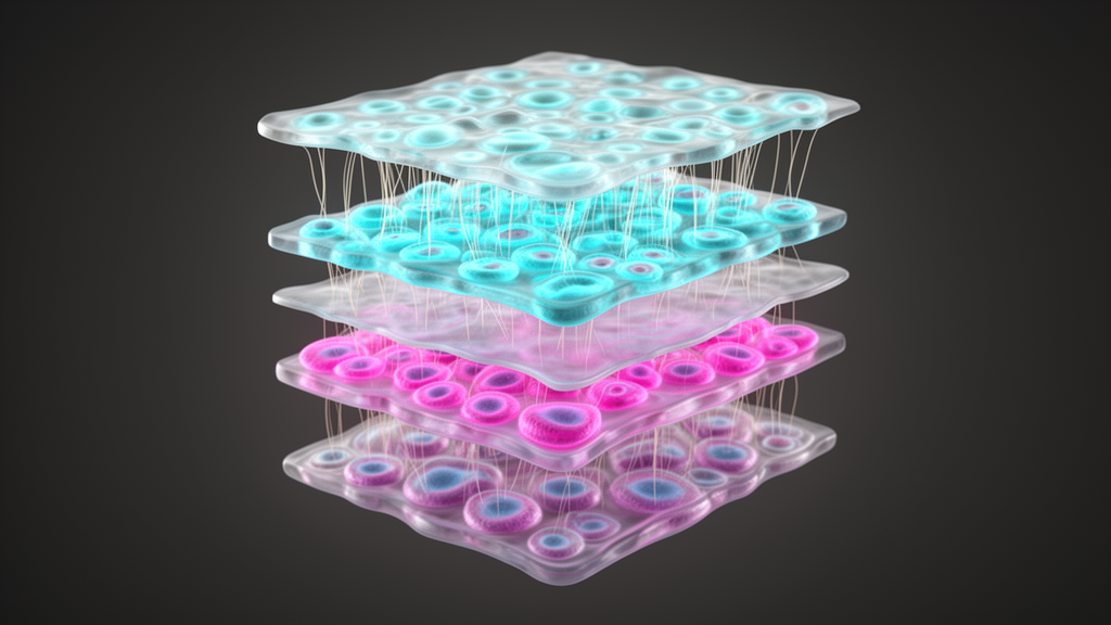
Constructing high-resolution three-dimensional tissue models from two-dimensional serial sections has emerged as a fundamental challenge in spatial biology, where linking cellular architecture to macroscopic organ function requires tracking millions of individual cells across consecutive physical slices. To resolve these spatial discrepancies, researchers developed Space-map, an automated computational framework designed to assemble coherent three-dimensional tissue architectures from massive single-cell coordinate datasets. The method unites two-dimensional cell-density rasterization, hybrid multimodal feature matching, and large deformation diffeomorphic metric mapping to automate global coordinate alignment across serial sections while safeguarding fine microanatomical boundaries. Benchmarking across diverse tissue systems demonstrated that the platform delivers substantially higher registration accuracy alongside up to a tenfold acceleration in processing speed compared with existing registration algorithms, establishing a scalable foundation for constructing human organ atlases at true single-cell resolution.
The mathematical and computational foundation of Space-map addresses the steep scalability bottlenecks inherent to single-cell spatial profiling technologies. When tissue sections contain hundreds of thousands or millions of measured cells, traditional pairwise registration algorithms that calculate interactions directly between individual cell coordinates rapidly become computationally intractable. Space-map circumvents this challenge by implementing a dimensionality reduction strategy that rasterizes discrete cell coordinates into continuous two-dimensional cell-density maps. Given a collection of two-dimensional cell coordinates, the algorithm determines the maximum bounding scale and divides the area using a predefined scaling factor to generate a uniform grid. Each grid cell records the count of falling cell centroids, transforming discrete point clouds into structured density images. This formulation reduces the core computational complexity from quadratic scaling to linear scaling relative to cell number.
Once density fields are generated and normalized within a standardized coordinate framework, Space-map executes a robust coarse alignment protocol engineered to overcome section tearing, missing tissue fragments, and major rotational displacements. The algorithm extracts salient anatomical landmarks by combining the traditional Scale-Invariant Feature Transform with the Transformer-based LoFTR algorithm, creating a comprehensive pool of complementary local and contextual feature points. To prevent spatial overfitting in dense tissue regions, the method enforces a feature-filtering constraint that retains only a single feature point per grid unit by default. Space-map then applies an iterative two-tier optimization scheme based on random sample consensus to compute global affine transformations encompassing rotation, scaling, and translation. In each iteration, high-scoring candidate feature pairs are sampled to fit transformation matrices, and the optimal alignment is selected by maximizing the Dice overlap coefficient between transformed sections. Rather than treating section pairs in isolation, this coarse stage considers preceding aligned sections to preserve cumulative structural continuity and prevent rotational drift across extended serial stacks.
Following global affine alignment, Space-map resolves complex local nonlinear tissue deformations using Large Deformation Diffeomorphic Metric Mapping. The diffeomorphic refinement stage models these local distortions by integrating smooth, time-varying velocity fields through continuous energy minimization. Because diffeomorphic mappings are inherently smooth, invertible, and topology-preserving, this mathematical formulation prevents artificial tissue folding, boundary tearing, or coordinate inversion. The optimization employs a multiresolution schedule alongside a center-reference alignment strategy, progressively establishing complete deformation field sequences across adjacent layers. By coupling multiscale feature detection with topology-preserving velocity fields, the algorithm balances rigid global alignment with localized structural compliance, ensuring that microscopic tissue compartments remain spatially coherent across physical gaps.
The research team demonstrated the experimental capabilities of Space-map by constructing a high-resolution three-dimensional atlas of a human colon polyp profiled with the imaging-based Xenium spatial transcriptomics platform. The biological specimen, representing a tubular adenoma with low-grade dysplasia, was collected during colectomy from a twenty-three-year-old male patient diagnosed with familial adenomatous polyposis under Stanford Institutional Review Board approval. The tissue block was formalin-fixed, paraffin-embedded, and sliced into thirty continuous serial sections cut at a uniform thickness of five micrometers. Profiling was conducted using a targeted four-hundred-twenty-two-gene panel combining a predesigned colon panel with custom disease-associated markers, yielding a dataset comprising 2.7 million cells and 123.6 million individual transcript localizations. Each physical section captured an average of ninety-three thousand cells, forty-eight transcripts per cell, and twenty-six distinct genes per cell, spanning thirty-nine annotated cell types that captured healthy epithelial hierarchies, dysplastic states, and infiltrating stromal and immune compartments.
Detailed histological inspection of the serial transcriptomic stack revealed a substantial physical discontinuity between sections twenty-two and twenty-three, which histopathology consultation identified as an approximate thirty-micrometer tissue loss incurred during ribbon trimming. Space-map was consequently deployed across the continuous twenty-section series spanning sections three through twenty-two to reconstruct the finalized three-dimensional polyp model. The resulting digital volume accurately resolved the tortuous microarchitecture of dysplastic intestinal crypts alongside localized cellular neighborhoods across tissue depth. In three dimensions, distinct cell types occupied characteristic spatial niches: myofibroblasts populated the supporting stroma, cytotoxic CD8-positive T lymphocytes exhibited diffuse infiltrative patterns, stem-like progenitor cells clustered in localized niches, and immature enterocytes formed continuous epithelial sheaths. Complementary hematoxylin and eosin brightfield histology images obtained from the identical slides were co-registered using the derived coordinate transformations, demonstrating the platform’s capacity to integrate histological morphology with cellular spatial transcriptomics.
To evaluate whether Space-map generalized across analytical modalities and healthy organ systems, the investigators reconstructed three-dimensional reference volumes from multiplexed spatial proteomics data acquired with the CODEX assay. Formalin-fixed paraffin-embedded tissue cores encompassing full-thickness intestinal walls were obtained from the normal sigmoid colon and duodenum of a fifty-six-year-old deceased male organ donor. Serial sections cut at five-micrometer intervals were stained with a fifty-two-protein antibody panel targeting epithelial, stromal, vascular, lymphatic, and immune cell lineages. Each section yielded an average of one hundred ninety-six thousand cells in the duodenum and one hundred six thousand cells in the sigmoid colon. Across sixteen aligned serial sections spanning an eighty-micrometer physical depth, Space-map reconstructed complete three-dimensional tissue blocks containing 3.1 million cells for the duodenum and 1.7 million cells for the sigmoid colon, successfully recapitulating mucosal folding, submucosal boundaries, and muscularis layers.
Visual and quantitative inspection of the reconstructed intestinal volumes revealed contrasting structural organization between healthy bowel compartments and the precancerous adenoma. The healthy sigmoid colon model preserved discrete, organized crypt columns, well-defined lamina propria spacing, and organized lymphoid follicles enriched for B lymphocytes, whereas the duodenal volume captured continuous mucosal surfaces and villus-like projections. In contrast to the orderly compartmentalization of the normal intestine, the adenomatous polyp exhibited partially intermingled spatial domains along the z-axis, where epithelial, stromal, immune, and neural cell populations formed extended irregular territories. Quantitative axial continuity analyses confirmed that multisection global alignment maintained cohesive glandular lumens and continuous core columns across serial sections. Conversely, naive pairwise-only registration produced localized blurring, artificial merging of adjacent crypt structures, and fragmented luminal pathways, highlighting the necessity of globally constrained registration across serial stacks.
Rigorous comparative benchmarking against established spatial registration tools, including PASTE and an adapted multi-section variant of STalign termed STalign-GT, confirmed the performance advantages of Space-map across multiple biological datasets. When evaluated on the Xenium polyp, CODEX sigmoid colon, and a public 128-section MERFISH mouse brain atlas, Space-map achieved the highest overall registration fidelity, yielding an average sparse Dice coefficient of 0.96 compared with 0.93 for STalign-GT and 0.90 for PASTE. The downsampled PASTE reconstructions exhibited substantial inter-section displacement and structural inversions, reflecting the severe memory and computational constraints that prevented it from processing full-scale datasets. STalign-GT produced localized surface artifacts, uneven regions, and reduced crypt continuity, largely stemming from its reliance on manual landmark initialization. In contrast, Space-map achieved fully automated initialization through grid-based feature detection, improved structural similarity scores against common coordinate frameworks by twelve percent, and elevated neighboring cell-type matching accuracy by eight percent over STalign-GT.
Beyond registration accuracy, computational efficiency metrics demonstrated substantial practical advantages for high-throughput laboratory deployments. Space-map operated approximately tenfold faster than STalign-GT while consuming substantially less physical memory, requiring 1.6 gigabytes of random-access memory compared with 4.6 gigabytes for STalign-GT on matched tasks. When aligning dataset pairs containing between 187,000 and 1 million cells per section on a standard Apple M4 MacBook Air equipped with 16 gigabytes of RAM, Space-map completed registration within two to five minutes while maintaining memory utilization below 3.6 gigabytes. The platform successfully scaled to reconstruct datasets containing more than 10 million total cells distributed across thirty serial sections. This marked operational efficiency stems directly from decoupling the registration runtime from total cell numbers through rasterized density representations, ensuring that computational overhead scales primarily with grid pixel dimensions rather than cell counts.
Despite its demonstrated registration fidelity and scalability, the authors delineated specific operational boundaries and technical constraints inherent to the Space-map framework. Because cell positions are mapped onto discrete grids, fine-scale spatial relationships that fall below the selected grid resolution cannot be directly constrained during the initial alignment phase, necessitating higher grid resolutions or adaptive feature budgets when analyzing highly disordered, densely packed tumors. Furthermore, while Space-map tolerates moderate section spacing and missing tissue gaps, severe mechanical damage, substantial tissue loss, or highly irregular physical spacing can compromise non-rigid diffeomorphic matching. To prevent algorithmic overfitting in poorly matched regions, the software includes configurable modes that permit users to prioritize global shape preservation over local similarity matching. The method has been validated primarily on continuous serial sections spaced at five micrometers, meaning that application to thick sections exceeding one hundred micrometers imaged via light-sheet fluorescence microscopy will require specialized surface-matching adaptations that remain untested on real-world specimens.
Subject of Research: Computational reconstruction of three-dimensional single-cell tissue atlases from serial histology sections
Article Title: Space-map: 3D single-cell atlas reconstruction from serial sections
Article References: Han, R., Ruan, C., Zhao, B., Tan, Y., Monte, E., Wei, B., Bi, J., Karathanos, T. V., Laquindanum, R., Charville, G. W., Lin, Y., Ling, N., Ford, J. M., Nolan, G. P., Wang, M., Zhu, C., & Snyder, M. P. (2026). Space-map: 3D single-cell atlas reconstruction from serial sections. Nature Methods. https://doi.org/10.1038/s41592-026-03310-8
Image Credits: AI Generated
DOI: 10.1038/s41592-026-03310-8
Keywords: Space-map, spatial transcriptomics, spatial proteomics, 3D tissue atlas, single-cell analysis, Xenium, CODEX, LDDMM, diffeomorphic mapping, computational biology, image registration, colon polyp, familial adenomatous polyposis, intestinal architecture, tissue morphology, structural biology
News Source: Gregory Coleman. (November 19, 2026). Space-map Reconstructs Three-Dimensional Single-Cell Tissue Atlases from Serial Sections. Scienmag.