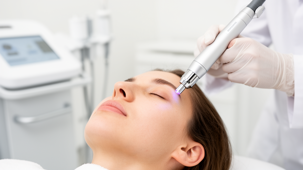
A randomized, split-face clinical trial conducted by dermatological researchers at Shanghai Ninth People’s Hospital has revealed that delivering 755-nanometer picosecond alexandrite laser energy through a uniform flat optic handpiece achieves long-term facial rejuvenation and pigment reduction comparable to a diffractive lens array while markedly reducing treatment discomfort and adverse inflammatory events. Investigating seventy-eight Chinese women presenting with symmetrical moderate facial photoaging alongside concurrent melasma, the researchers demonstrated that while both optical configurations significantly improved cutaneous appearance across twenty-four months of post-treatment monitoring, the flat optic beam yielded a 35.9 percent incidence of post-inflammatory hyperpigmentation compared to 60.3 percent on facial halves exposed to the diffractive lens array. Furthermore, the flat optic configuration facilitated a significantly more rapid initial clearance of pigmentary dyschromia and melasma severity across early treatment intervals, presenting clinicians with an evidence-based approach to mitigating hypermelanosis risks when managing complex photoaging in sensitive, pigment-dense Fitzpatrick skin types III and IV.
Treating facial photoaging in East Asian populations has historically posed substantial clinical hurdles because structural collagen degradation, solar elastosis, and coarsened skin texture frequently coexist with hyperactive epidermal and dermal melanogenesis manifested as melasma. Standard dermatological resurfacing modalities, particularly conventional ablative and non-ablative photothermal lasers, carry a well-documented risk of triggering post-inflammatory hyperpigmentation and aggravating underlying melasma patches due to collateral thermal injury and subsequent cutaneous inflammation. While the emergence of 755-nanometer picosecond alexandrite systems introduced ultra-short pulse durations that operate primarily via photomechanical stress waves rather than photothermal tissue destruction, clinical uncertainty has persisted regarding the comparative therapeutic profiles of its two primary delivery handpieces: the non-fractionated flat optic, which emits a homogeneous beam across the target area, and the diffractive lens array, which fractionates incident light into concentrated microbeams designed to initiate localized laser-induced optical breakdown within the epidermal stratum spinosum.
To rigorously evaluate the clinical and structural differences between these two optical modalities, investigators organized a prospective single-center trial spanning from August 2022 to August 2025 under protocol approval from the Medical Ethical Committee of Shanghai Ninth People’s Hospital and formal registration with the Chinese Clinical Trial Registry under identifier ChiCTR2200065607. From an initial screening cohort of eighty-five individuals, eighty adult female participants aged twenty to sixty years displaying symmetrical bilateral moderate photoaging and concurrent melasma were enrolled following written informed consent. Two participants subsequently discontinued participation due to international relocation, leaving seventy-eight patients with a mean age of 42.95 ± 8.19 years who completed the entire treatment sequence and longitudinal surveillance. Seventeen participants presented with Fitzpatrick skin phototype III, whereas sixty-one exhibited phototype IV, with the overall cohort exhibiting a mean duration since melasma diagnosis of 4.16 ± 2.23 years and well-balanced baseline photoaging severity scores across bilateral facial zones.
The study protocol utilized a randomized 1:1 split-face allocation structure designed to eliminate inter-individual physiological variability by applying one delivery mode to each hemiface. Randomization sequences generated by an independent statistician were concealed inside sequentially numbered opaque envelopes until revealed to the treating physician immediately prior to the inaugural session, resulting in thirty-eight participants receiving flat optic therapy on the left hemiface and forty on the right hemiface, with the contralateral side receiving diffractive lens array therapy. Both the participants and the independent panel of assessing dermatologists were blinded to the specific allocation scheme throughout all evaluation periods. Pre-treatment baseline clinical scores established that there were no statistically significant hemispheric disparities in baseline dyschromia, laxity, rhytids, or melasma severity, ensuring that observed post-intervention variances could be attributed directly to the differential performance of the optical delivery configurations under their established clinical operating parameters.
All laser treatments were executed by a single experienced practitioner using a commercial 755-nanometer picosecond alexandrite laser system configured with uniform core operational parameters across both facial halves, including a nominal fluence of 0.71 joules per square centimeter, a six-millimeter spot diameter, a pulse duration of 750 picoseconds, and a repetition frequency of 10 hertz. The flat optic handpiece delivered 2,000 to 2,500 pulses across three to four passes, whereas the diffractive lens array delivered 1,500 to 1,800 pulses across one to three passes, with passes adjusted to attain a clinical endpoint of mild uniform erythema devoid of tissue whitening or pinpoint hemorrhage. Treatments occurred over six scheduled sessions spaced four weeks apart, with standardized post-procedure ice application for fifteen minutes. Participants maintained strict daily photoprotection using broad-spectrum sunscreens rated at SPF 50+ while refraining from external retinoids, depigmenting topicals, chemical peelings, and alternative light-based therapies throughout the twenty-four-month trial duration.
Clinical efficacy was systematically recorded under standardized cross-polarized, parallel-polarized, and ultraviolet digital photography using dedicated clinical imaging hardware at baseline, four weeks following the first, third, and sixth treatments, and at a final twenty-four-month post-treatment evaluation visit. The trial’s designated primary outcome was the mean change in dyschromia scores from baseline to four weeks following the third treatment session, assessed on a validated four-point quartile photoaging scale by three blinded board-certified dermatologists whose evaluations demonstrated high inter-rater concordance with an intraclass correlation coefficient of 0.91. Secondary outcomes encompassed longitudinal changes in facial laxity, rhytid depth, skin texture, melasma extent graded through the hemi-Melasma Area and Severity Index, patient satisfaction tracked via five-point Likert scales, standardized visual analog scale pain ratings, blinded adverse event monitoring, and histometric quantification from an exploratory sub-study of consenting participants.
Statistical evaluation through linear mixed-effects models demonstrated that while both modalities produced highly significant longitudinal reductions in dyschromia over the entire experimental timeframe, the overall time-by-treatment interaction for dyschromia did not reach statistical significance, reflecting equivalent end-of-study pigmentary clearance. Dyschromia ratings declined from baseline values of 2.54 for the flat optic and 2.56 for the diffractive lens array to 1.60 and 1.65, respectively, following six sessions, before stabilizing at 1.79 and 1.86 at the twenty-four-month milestone. However, post-hoc pairwise analyses revealed that the flat optic mode achieved a significantly faster initial clearing trajectory during early intervention. Similarly, analysis of melasma severity demonstrated a statistically significant time-by-treatment interaction, with flat optic treatment producing superior early reductions in hemi-MASI scores by the third treatment session before both arms converged to comparable values of 3.76 for flat optic and 3.82 for diffractive delivery after the complete six-session protocol.
In contrast to the pigmentary outcomes, linear mixed modeling of cutaneous surface texture demonstrated a statistically significant time-by-treatment interaction that favored the diffractive lens array during initial sessions. Post-hoc tests established that the diffractive lens array elicited significantly greater texture refinement after a single treatment session, lowering texture scores from a baseline of 2.31 to 2.03, compared to a reduction from 2.28 to 2.18 observed with the flat optic handpiece. By the completion of six sessions, texture ratings stood at 1.948 on flat optic sides and 1.85 on diffractive sides, subsequently settling at 2.03 and 2.05 at twenty-four months. Evaluations of skin laxity and facial rhytids showed significant time-dependent enhancements across both modalities, but lacked statistically significant time-by-treatment interaction effects over the two-year timeline, confirming that long-term structural tissue tightening and wrinkle attenuation remained fundamentally equivalent between the two optical beam configurations.
The safety and tolerability metrics exhibited pronounced, clinically meaningful disparities between the two delivery approaches, heavily favoring the flat optic modality among the cohort. Pain scores tabulated across all six sessions via a ten-point visual analog scale revealed significantly lower intra-procedural discomfort on hemifaces treated with the flat optic, averaging 4.18 compared to 5.83 on contralateral sides treated with the diffractive lens array. Furthermore, paired nominal analysis using McNemar’s test confirmed a statistically significant divergence in post-inflammatory hyperpigmentation incidence, which occurred on twenty-eight flat optic hemifaces versus forty-seven diffractive lens array hemifaces. Among those developing post-inflammatory hyperpigmentation, 60.7 percent of flat-optic-treated areas resolved within one single week, whereas 27.7 percent of diffractive-treated areas suffered prolonged pigmentation lasting longer than four weeks, with two individuals exhibiting persistent dyschromia beyond twelve weeks.
Patient-reported downtime further corroborated the gentler physical profile of the flat optic handpiece, showing significantly shorter average durations for post-treatment erythema, edema, and cutaneous sensitivity. Specifically, erythema persisted for an average of 2.51 days following flat optic application compared to 4.59 days after diffractive lens array exposure, facial edema resolved in 1.24 days versus 1.67 days, and post-procedure skin sensitivity subsided in 1.74 days versus 3.77 days. The duration of post-laser cutaneous dryness exhibited a modest numerical difference of 2.40 days versus 2.94 days that did not attain statistical significance. A single participant in the diffractive cohort developed transient epidermal vesiculation that cleared spontaneously within forty-eight hours without scarring. Despite these differing adverse profiles, overall patient satisfaction assessed at one month post-treatment was exceptionally high in both groups, recording 94.9 percent satisfaction for the flat optic and 93.6 percent for the diffractive lens array.
Microscopic insights obtained from an exploratory histological sub-study involving twelve bilateral punch biopsy specimens taken from the nasolabial folds of three participants reinforced the clinical finding of comparable long-term structural remodeling. Quantitative analysis demonstrated that mean epidermal thickness increased significantly from baseline under both protocols, rising from 44.37 to 52.02 micrometers with the flat optic and from 47.04 to 59.15 micrometers with the diffractive lens array, without a statistically significant difference between modalities. Immunohistochemical H-score evaluations demonstrated significant enhancements in dermal neocollagenesis, type III collagen synthesis, mature elastin fiber density, and tropoelastin expression across both treatment arms four weeks following the third session. Although the diffractive lens array achieved a statistically significant rise in type I collagen deposition that was only a trend in the flat optic arm, inter-treatment comparisons showed no statistically significant differences across any collagenous or elastic matrix markers.
The investigators emphasized several methodological constraints, noting that the exploratory histological findings reflect a very small cohort of three biopsied participants and must be considered hypothesis-generating descriptive data rather than definitive proof of biological equivalence. Additionally, while the primary dyschromia endpoint was strictly confirmatory, secondary clinical photoaging comparisons were not formally corrected for multiplicity, and minor differences in post-procedure erythema could theoretically have influenced clinical assessor masking. Most importantly, because the two delivery modalities operated under standardized clinical pulse and pass protocols rather than energy-matched parameters, observed outcomes reflect protocol-level differences rather than isolated optical geometry alone. Nevertheless, the trial confirms that flat optic 755-nanometer picosecond alexandrite laser protocols deliver equivalent durable facial rejuvenation while substantially lowering post-inflammatory hyperpigmentation risks, offering a highly advantageous therapeutic strategy for managing photoaging complicated by melasma in Asian skin.
Subject of Research: Comparative efficacy and safety of flat optic versus diffractive lens array picosecond lasers for facial rejuvenation in women with photoaging and melasma
Article Title: Flat optic versus diffractive lens array for 755-nm picosecond laser treatment of melasma-associated photoaging: a randomized split-face trial in Chinese women
Article References: Sun, Q., Liu, Y., Cen, Q., Han, Y., Shang, Y., Ma, G., & Wu, P. (2026). Flat optic versus diffractive lens array for 755-nm picosecond laser treatment of melasma-associated photoaging: a randomized split-face trial in Chinese women. Frontiers in Medicine, 13, Article 2007032. https://doi.org/10.3389/fmed.2026.2007032
Image Credits: AI Generated
DOI: 10.3389/fmed.2026.2007032
Keywords: picosecond laser, alexandrite laser, melasma, photoaging, facial rejuvenation, post-inflammatory hyperpigmentation, split-face trial, flat optic, diffractive lens array, dermatology, Fitzpatrick skin phototypes, dyschromia, collagen remodeling, skin laxity, elastin, hemi-MASI, aesthetic medicine
News Source: Ophelia Keating. (November 18, 2026). Flat Optic Picosecond Laser Cuts Pigmentation Risks in Split-Face Trial. Scienmag.