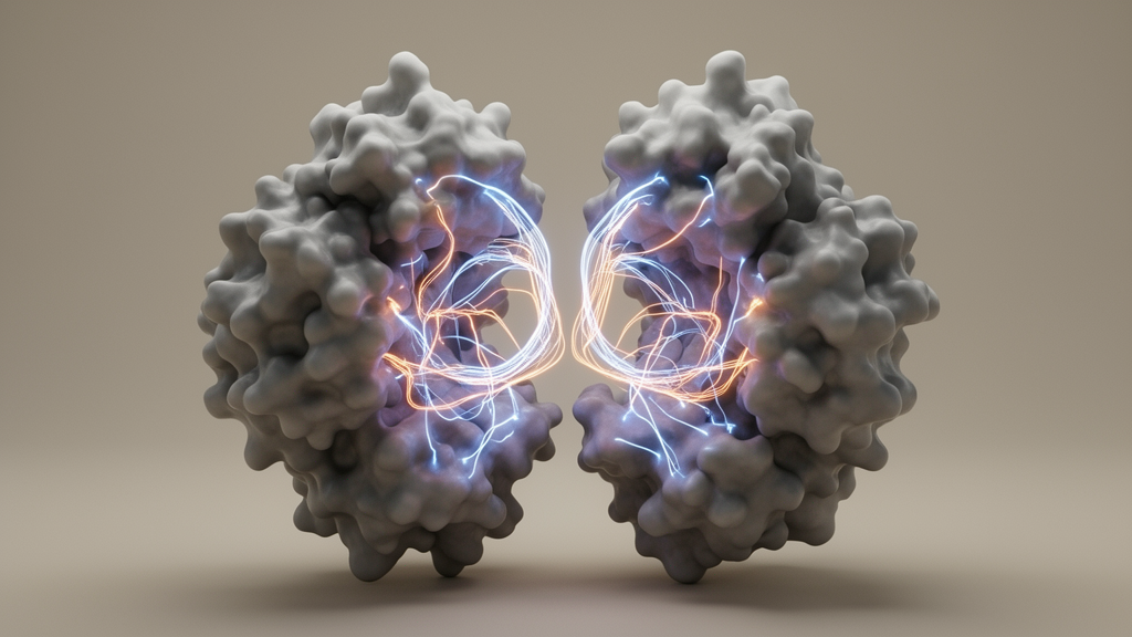
Mammalian nitric oxide synthases synthesize nitric oxide, an indispensable signaling molecule that regulates neurotransmission, cardiovascular tone, and immune defenses against cellular pathogens. When enzymatic activity is disrupted or aberrantly controlled, the resulting imbalance directly contributes to severe clinical pathologies, including neurodegenerative decline, septic shock, and ischemic tissue damage. Nitric oxide synthase catalysis depends upon an intricate cascade of electron transfers across five essential cofactors, where electrons supplied by an NADPH-dependent reductase domain pass via flavin adenine dinucleotide and flavin mononucleotide subdomains to an oxygenase domain containing a catalytic heme center and a tetrahydrobiopterin cofactor that converts L-arginine into nitric oxide. Although this fundamental biological pathway has been recognized for decades, the complete structural basis of human nitric oxide synthase function has remained elusive because of the extreme conformational flexibility and multi-subunit architecture of the complex. Reporting in Nature Structural & Molecular Biology, a research team led by Kanghyun Lee and Daniel R. Southworth resolved high-resolution cryo-electron microscopy structures of the intact human inducible nitric oxide synthase holoenzyme bound to calmodulin, demonstrating that the enzyme operates in an engaged, asymmetric conformation wherein a single reductase arm crosses between subunits to deliver electrons in trans.
To capture this transient and highly dynamic assembly, the investigators reconstituted and purified the full-length human inducible nitric oxide synthase complex coexpressed with human calmodulin in Escherichia coli BL21(AI) cells. Unlike previous structural predictions generated by computational tools such as AlphaFold3, which modeled the full-length tetramer in an inactive, extended state with the flavin mononucleotide and heme cofactors separated by more than 80 angstroms, the experimental complex required specialized stabilization to resolve active functional intermediates. The researchers isolated the CaM-bound dimer by size-exclusion chromatography, verifying a molecular mass of approximately 300 kilodaltons with multiangle light scattering. Functional integrity was rigorously confirmed before imaging, showing efficient heme incorporation of approximately 2.6 nanomoles of P450 per milligram of protein and robust nitric oxide synthesis rates of approximately 94 nanomoles of nitric oxide per minute per nanomole of P450 through carbon monoxide binding and oxyhemoglobin oxidation assays. Because preliminary uncrosslinked two-dimensional class averages showed well-resolved density exclusively for the oxygenase dimer while the reductase and calmodulin regions remained blurred by severe interdomain motion, the team introduced the chemical crosslinker disuccinimidyl dibutyric acid to trap functional interactions prior to vitrification.
Cryo-electron microscopy data collection across 35,896 movie micrographs on Titan Krios transmission electron microscopes operating at 300 kilovolts incorporated specimen tilting and non-ionic detergent supplementation with DDM, octyl beta-D-glucopyranoside, and amphipol to overcome severe particle orientation bias. Extensive three-dimensional classification and heterogeneous refinement separated the particle population into two dominant structural architectures. The predominant class, comprising roughly 35 percent of the dataset, refined to a 2.6-angstrom resolution structure representing an isolated oxygenase dimer with only weak, flexibly associated calmodulin density, closely matching previously determined murine crystal structures with an alpha-carbon root-mean-square deviation of 0.5 angstroms across 409 pruned atom pairs. The second major class, containing 21 percent of the particle images, refined to a 3.0-angstrom consensus reconstruction representing an engaged holoenzyme state. In this engaged assembly, the oxygenase dimer achieved 2.5-angstrom local resolution, while the flexibly anchored reductase and calmodulin arm resolved at 4.5 to 6.5 angstroms, demonstrating that only one reductase arm docks stably onto the oxygenase platform at any single moment while the opposing arm remains flexibly disengaged.
Within this engaged architecture, the reductase domain adopts an unprecedented deshielded output conformation that differs radically from the shielded resting states observed in crystallographic studies of isolated neuronal nitric oxide synthase. In the canonical shielded conformation, the flavin mononucleotide subdomain remains tightly docked against the flavin adenine dinucleotide-binding ferredoxin-NADP+ reductase subdomain, maintaining a cofactor separation of only 3.4 angstroms that favors interflavin reduction but blocks external electron egress. By aligning the ferredoxin-NADP+ reductase cores, the researchers discovered that the flavin mononucleotide subdomain in the engaged inducible nitric oxide synthase complex rotates by approximately 86 to 90 degrees relative to its shielded position. This substantial domain reorientation widens the distance between the two flavin cofactors to 10.3 angstroms while turning the isoalloxazine ring such that its C7 and C8 methyl substituents point outward toward the opposing monomer’s catalytic oxygenase cleft, effectively converting the domain into an electron-donating output state.
This massive subdomain rotation is mechanically stabilized and guided by an extensive network of interdomain contacts involving calmodulin and the interdomain hinge region. In the intact complex, calmodulin forms a critical salt bridge between its glutamate 48 residue and arginine 536 of inducible nitric oxide synthase at the junction connecting the calmodulin-binding helix to the flavin mononucleotide subdomain, alongside a secondary stabilizing contact between aspartate 51 of calmodulin and arginine 687. Comparison with prior isolated crystal structures showed that calmodulin binding causes an additional 6-degree tilt of the calmodulin-binding helix, displacing the flavin cofactor by roughly 15 angstroms closer to the oxygenase active site. Simultaneously, an interface formed between a helical turn linking beta strands 5 and 6 and helix 7 of the flavin mononucleotide subdomain establishes a hydrophobic contact pocket with proline 865 of the ferredoxin-NADP+ reductase subdomain via threonine 654 and proline 655, further locked by a hydrogen bond between glutamine 840 and glutamine 675. Computational docking revealed that the canonical shielded state cannot physically assemble while calmodulin remains bound because steric clashes between the ferredoxin-NADP+ reductase beta-hairpin finger and the calmodulin C-lobe prevent simultaneous occupancy.
To determine whether calmodulin binding alone induces flavin mononucleotide subdomain deshielding in the absence of an intact catalytic partner, the researchers examined isolated monomeric inducible nitric oxide synthase fractions lacking heme incorporation using single-particle cryo-electron microscopy across 6,855 videos. Three-dimensional classifications of this uncrosslinked monomer revealed that the disengaged reductase arm samples both a shielded state at 4 to 5 angstroms resolution and a rotated, deshielded conformation. To corroborate these interdomain geometries in the full holoenzyme, the authors performed chemical crosslinking mass spectrometry with disuccinimidyl dibutyric acid using parallel accumulation-serial fragmentation data-dependent acquisition followed by targeted parallel reaction monitoring on a timsTOF fleX mass spectrometer. This analysis identified 60 high-confidence crosslinks, including 17 intermolecular calmodulin-synthase pairs and 43 intramolecular connections. Fifty of these crosslinks satisfied the maximum 40-angstrom physical distance constraint imposed by the crosslinker, with specific crosslinks between calmodulin lysine 95 and oxygenase lysine 88, as well as connections linking the second oxygenase monomer to the flavin mononucleotide subdomain, directly validating the engaged quaternary arrangement.
Structural dissection of the engaged complex identified four discrete interprotomer and interdomain interaction sites responsible for docking the reductase arm across the oxygenase dimer surface. The first interface involves basic residues arginine 83 and histidine 84 on the second oxygenase monomer, which engage a calcium-coordinating loop in calmodulin containing aspartate 132 and aspartate 134. The second site comprises acidic residues on calmodulin helix 7, specifically aspartate 119, glutamate 120, and aspartate 123, which form complementary electrostatic networks across the dimer interface with arginine 86 and lysine 88 on the second monomer and lysine 497 on the first monomer. The third interface tethers the ferredoxin-NADP+ reductase core in cis to the first oxygenase domain through a cation-pi interaction between tyrosine 812 at the tip of the reductase beta-hairpin finger and oxygenase arginine 398, accompanied by hydrophobic contacts through valine 814 and an adjacent salt bridge between lysine 817 and glutamate 402. The fourth interface positions the flavin mononucleotide subdomain directly against a unique helix-turn-helix motif on the second oxygenase monomer via electrostatic interactions between aspartate 573, lysine 574, and arginine 576 and oxygenase residues lysine 155 and glutamate 156.
The biological requirement for these structural contact points was systematically evaluated by site-directed mutagenesis and quantitative enzymatic activity measurements standardized against P450 content. Alanine substitution of the distal calmodulin-docking site through an R83A and H84A double mutant decreased catalytic nitric oxide generation by approximately 25 percent with a statistically significant difference of P equals 0.00109. Alanine substitution of the acidic calmodulin triad comprising D119A, E120A, and D123A at the central dimer junction nearly abolished enzymatic activity, confirming that this electrostatic bridge is critical for functional reductase engagement. Demonstrating the specificity of this contact, individual charge-reversal mutations in the CaM(E120K) or Oxy(K497E) pair reduced catalytic output by approximately half, whereas combining both charge reversals in a reciprocal double mutant restored catalytic turnover to wild-type levels with a non-significant P value of 0.0994. Similarly, disrupting the cis beta-hairpin finger via a Y812A mutation reduced synthesis rates by nearly half, while charge reversal at the helix-turn-helix motif through an E156R mutation caused a 25 percent activity decline with P equals 0.0102, demonstrating that multiple cooperative interfaces stabilize the active holoenzyme.
Because the reductase arm retained measurable flexibility relative to the oxygenase platform even within the crosslinked engaged state, the investigators applied three-dimensional variability analysis to interrogate conformational dynamics across the particle population. This analysis resolved two distinct functional end states within the engaged landscape, designated as distal and proximal conformations, each refined to approximately 3.0 angstroms resolution. Comparing these two states demonstrated that while the oxygenase dimer remains rigid, the ferredoxin-NADP+ reductase, flavin mononucleotide, and calmodulin domains undergo coordinated spatial displacements exceeding 10 to 12 angstroms in root-mean-square deviation. In the distal conformation, the flavin mononucleotide cofactor resides 11.4 angstroms from the flavin adenine dinucleotide group, 13.4 angstroms from the oxygenase heme iron, and 10.3 angstroms from a conserved catalytic tryptophan residue, tryptophan 372. This geometry places the flavin intermediate closer to the primary electron input site, likely optimizing electron transfer from flavin adenine dinucleotide to flavin mononucleotide.
In the corresponding proximal conformation, the flavin mononucleotide subdomain slides closer against the second oxygenase subunit, narrowing the distance between the flavin mononucleotide cofactor and the catalytic heme iron to 9.1 angstroms. Crucially, the separation between the flavin mononucleotide methyl substituents and the indole sidechain of tryptophan 372 contracts to just 5.3 angstroms, creating an optimal low-barrier pathway for rapid electron injection into the oxygenase active site. Conserved aromatic residues, including tyrosine 631 and proline 632 in the reductase domain along with methionine 434 and tryptophan 463 near the heme, align into a continuous tunneling conduit. Tetrahydrobiopterin exhibits continuous electron density with tryptophan 463 and extends toward arginine 199 and arginine 381, stabilizing the redox cofactor during oxyferrous heme activation. These observations demonstrate that human inducible nitric oxide synthase does not require continuous large-scale dissociation into fully shielded conformations between catalytic steps. Instead, persistent calmodulin tethering holds the reductase arm in an engaged trans orientation, allowing rapid conformational shuttling between distal flavin-charging and proximal heme-reducing states to drive sustained enzymatic synthesis.
Subject of Research: Structural basis of human inducible nitric oxide synthase holoenzyme function and interdomain electron transfer
Article Title: Structural basis of electron transfer by the human nitric oxide synthase holoenzyme complex
Article References: Lee, K., Martinez-Ramos, C., Jiang, T., Pospiech, T. H., Jr., Tse, E., Lau, M., Feng, C., Osawa, Y., & Southworth, D. R. (2026). Structural basis of electron transfer by the human nitric oxide synthase holoenzyme complex. Nature Structural & Molecular Biology. https://doi.org/10.1038/s41594-026-01939-9
Image Credits: AI Generated
DOI: 10.1038/s41594-026-01939-9
Keywords: nitric oxide synthase, cryo-electron microscopy, iNOS holoenzyme, calmodulin, electron transfer, FMN subdomain, heme cofactor, FAD cofactor, protein crosslinking, DSBU, mass spectrometry, conformational dynamics, reductase domain, oxygenase domain, 3D variability analysis, redox biology, structural biology
News Source: Jason Bradley. (November 18, 2026). Cryo-EM Uncovers How Human Nitric Oxide Synthase Coordinates Intermonomer Electron Transfer. Scienmag.