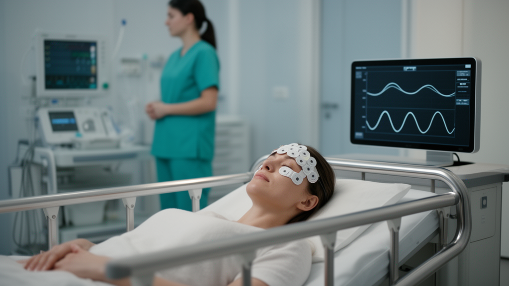
During surgical procedures requiring general anesthesia, clinical teams routinely rely on processed electroencephalography monitors to track brain activity and maintain appropriate depth of unconsciousness. These monitoring systems collect electrical signals from sensors placed across the forehead and compress complex neural oscillations into simplified numerical indices. However, clinical evidence increasingly indicates that relying strictly on single numerical depth targets fails to reliably prevent postoperative delirium in older surgical patients. Writing in the Journal of Clinical Monitoring and Computing, researcher Vikas Chauhan synthesized evidence from randomized clinical trials, systematic reviews, and neurophysiologic studies to evaluate the diagnostic utility of limited-channel frontal electroencephalography. The comprehensive narrative review evaluated research published through April 30, 2026, demonstrating that while titrating anesthetic agents to processed numerical targets alone does not prevent acute postoperative cognitive dysfunction, the underlying electrical waveforms provide crucial contextual data. The author identified four clinically accessible frontal brainwave patterns that illuminate individual variations in anesthetic sensitivity, baseline cerebral reserve, and recovery dynamics, offering an interpretive framework for perioperative monitoring.
Within current clinical terminology, delirium forms part of a broader spectrum designated as perioperative neurocognitive disorders, which also includes pre-existing cognitive impairment, delayed neurocognitive recovery diagnosed within thirty days of an operation, and sustained postoperative neurocognitive disorders lasting up to twelve months. This acute state is consistently associated with prolonged hospital admissions, delayed functional independence, lasting cognitive decline, and elevated patient mortality. Older individuals presenting for major surgery frequently exhibit overlapping vulnerabilities, including multimorbidity, polypharmacy, baseline sensory and cognitive deficits, and reduced physiologic reserve, which are clinically captured through standardized assessments of physical frailty.
However, large randomized controlled trials have challenged this straightforward mechanistic assumption. In the ENGAGES trial, which randomized 1,232 older adults to processed electroencephalography-guided anesthesia or usual care, actively reducing time spent in burst suppression failed to decrease postoperative delirium rates. Similarly, the BALANCED trial evaluated 6,644 patients aged sixty and older randomized to lighter versus deeper anesthetic targets, finding no difference in one-year mortality, although a delirium substudy noted delirium occurred in twenty-eight percent of deeper-targeted patients compared to nineteen percent of lighter-targeted individuals. Extending these observations to major cardiac surgery, the 2024 ENGAGES-Canada trial examined 1,140 older surgical patients, showing that despite achieving a sixty-six percent reduction in burst suppression through guided care, postoperative delirium developed in eighteen percent of participants across both study arms. Evidence syntheses also demonstrate regional heterogeneity, with pooled trial benefits concentrated in European studies but absent across North American cohorts, indicating that processed numerical guidance is not a uniform preventive intervention.
The discrepancy between numerical monitor readings and clinical neurocognitive outcomes stems largely from the severe computational data compression inherent in processed monitoring algorithms. A proprietary monitor calculates an index by summarizing complex biophysical activity into a single dimensionless number, meaning two distinct patients exhibiting identical numerical values can possess completely divergent underlying neurophysiologic states. In one patient, the number may reflect robust, organized alpha rhythms paired with steady slow-wave oscillations, whereas in another, the identical value may conceal severely attenuated alpha power, unstable spectral dynamics, and intermittent periods of electrical suppression. Furthermore, postoperative delirium is fundamentally a multifactorial syndrome that develops through complex interactions involving pre-existing biological susceptibility, systemic surgical inflammation, unmanaged pain, acute sleep disruption, sedative medications, hypothermia, anemia, and hemodynamic instability. Modifying anesthetic delivery to keep an algorithmic index within a prescribed window addresses only one component of this pathophysiologic cascade, rendering isolated numerical guidance insufficient for delirium prevention.
A central feature examined in the narrative review is frontal burst suppression, a neurophysiologic pattern defined by alternating intervals of high-amplitude electrical activity and periods of relative electrical quiescence. Burst suppression has traditionally served as a marker of profound pharmacologic cortical depression, severe hypothermia, metabolic encephalopathy, or acute cerebral injury. Multiple observational investigations and systematic reviews have established a robust association between intraoperative burst suppression duration and postoperative delirium. For example, meta-analyses in older surgical cohorts have documented an approximate forty-one percent higher relative risk of delirium when burst suppression is present, with delirium incidence reaching twenty-two percent in patients exhibiting suppression compared to thirteen percent in those without it. Furthermore, a comprehensive systematic review encompassing forty-two studies and more than 13,000 patients confirmed that both the presence and cumulative duration of burst suppression correlated with delirium even when anesthesia delivery was guided by processed monitoring. However, because randomized suppression-minimization protocols do not consistently prevent delirium, researchers conclude that burst suppression functions as an informative marker of baseline cerebral vulnerability rather than a direct, independent causal mechanism.
Building upon these mechanistic considerations, the author outlined four distinct, clinically accessible frontal electroencephalographic patterns that provide meaningful diagnostic context when interpreted alongside pharmacology and physiology. The first pattern is reduced frontal alpha power, where the 8 to 12 Hertz oscillations typically induced by drugs such as propofol appear attenuated or disorganized. In healthy younger brains, general anesthesia produces coherent anteriorization of alpha rhythms, but aging and impaired cognitive performance weaken this response. The extensive meta-analysis of over 13,000 patients demonstrated that lower intraoperative alpha mean power and decreased alpha peak frequency were among the electroencephalographic variables most consistently linked to delirium, establishing alpha attenuation as a subtle marker of diminished cognitive reserve. The second pattern involves early or disproportionate burst suppression, which occurs at unexpectedly low anesthetic concentrations or modest age-adjusted drug exposures. Research by Fritz and colleagues revealed that suppression triggered at lower volatile anesthetic concentrations predicted delirium more strongly than total suppression duration alone, highlighting individual pharmacodynamic sensitivity. The third pattern encompasses persistent spectral slowing and depressed spectral edge frequency, reflecting impaired cortical flexibility when slow-delta dominance continues uninterrupted. The fourth pattern consists of disorganized emergence trajectories, where patients transition toward consciousness with persistent delta activity, delayed alpha recovery, or erratic state changes rather than progressive spectral reorganization.
The clinical interpretation of these electroencephalographic signals requires strict alignment with the specific pharmacologic properties of administered anesthetic agents. General anesthetics produce drug-specific electroencephalographic signatures that cannot be aggregated into a single depth scale. Propofol administration characteristically induces coordinated slow-delta and frontal alpha oscillations at surgical levels of unconsciousness before progressing to burst suppression at higher concentrations. Volatile halogenated agents, such as sevoflurane and desflurane, generate dose-dependent slow-delta and theta activity accompanied by frontal alpha oscillations at lower concentrations, shifting toward suppression at elevated doses with spectral nuances distinct from propofol. Ketamine, an N-methyl-D-aspartate receptor antagonist, maintains high-frequency gamma oscillations rather than uniform cortical depression, while systemic opioids systematically attenuate high-frequency power.
Equally critical to electroencephalographic interpretation is the contemporaneous evaluation of patient hemodynamics, metabolic stability, and technical signal quality. The aging cerebral vasculature demonstrates heightened vulnerability to arterial hypotension, autoregulatory failure, hypocapnia, hypoxemia, and anemia, all of which compromise cerebral oxygen delivery. An unexpected decline in raw amplitude, sudden spectral slowing, or spontaneous burst suppression may stem directly from transient cerebral hypoperfusion rather than anesthetic overdose, requiring immediate blood pressure restoration or ventilation adjustments rather than drug titration. Concurrently, clinicians must maintain awareness of technical artifacts that distort processed algorithms. High-frequency electromyographic interference from facial muscle tension, shivering, surgical electrocautery, and external warming devices can artificially elevate processed index values, giving a false impression of inadequate depth. Reviewing raw waveforms and density spectral arrays enables clinicians to verify signal integrity before forming clinical judgments.
The review emphasizes that perioperative neurocognitive protection requires continuous cognitive monitoring across the entire surgical continuum, bridging preoperative assessment, intraoperative maintenance, emergence, and postoperative critical care. Rather than functioning as an isolated operating-room depth gauge, frontal monitoring provides longitudinal physiological insights that can inform care transitions. Preoperative cognitive screenings, such as the Montreal Cognitive Assessment or Mini-Cog, alongside documented physical frailty, establish baseline vulnerability before induction. During emergence, the failure to recover organized frontal alpha oscillations provides valuable prognostic data regarding recovery trajectories. Prospective studies have shown that the absence of dominant alpha oscillations during emergence from delta-dominant anesthesia directly predicts postoperative neurocognitive impairment. Communicating intraoperative findings—such as persistent suppression at modest drug doses or disorganized emergence—to postanesthesia care units and intensive care teams allows clinicians to implement targeted surveillance, avoid deliriogenic medications, support circadian sleep architecture, and apply structured interventions such as the multicomponent ABCDEF bundle.
Despite the clinical insights provided by limited-channel frontal monitoring, the author explicitly noted critical methodological limitations governing these observations. As a narrative review rather than a formal meta-analysis, the synthesis did not follow a predefined systematic protocol or calculate pooled effect estimates. The four identified electroencephalographic patterns represent clinical interpretive constructs derived from limited-channel frontal sensors rather than validated diagnostic categories or proven treatment endpoints. Because commercial frontal sensors sample only prefrontal and frontotemporal regions, they cannot evaluate posterior cortical activity, temporal lobe dynamics, or large-scale neural network connectivity; mechanistic concepts such as thalamocortical alpha anteriorization rest on separate high-density recordings and computational modeling. Furthermore, current evidence linking these frontal patterns to neurocognitive disorders remains largely observational and subject to confounding by patient age, drug exposure, and cardiovascular status. Prospective, randomized trials incorporating agent-specific analyses and multimodal physiological variables will be necessary before pattern-guided electroencephalography interpretation can be formally established as a validated clinical standard for delirium risk reduction.
Subject of Research: Frontal electroencephalography pattern interpretation and postoperative delirium risk in older surgical patients
Article Title: Beyond processed depth targets: frontal EEG patterns, brain vulnerability, and postoperative delirium in older adults
Article References: Chauhan, V. (2026). Beyond processed depth targets: frontal EEG patterns, brain vulnerability, and postoperative delirium in older adults. Journal of Clinical Monitoring and Computing. https://doi.org/10.1007/s10877-026-01507-y
Image Credits: AI Generated
DOI: 10.1007/s10877-026-01507-y
Keywords: electroencephalography, postoperative delirium, anesthesia monitoring, processed EEG, frontal alpha power, burst suppression, spectral slowing, spectral edge frequency, emergence trajectories, older adults, cerebral reserve, brain vulnerability, perioperative neurocognitive disorders, bispectral index, propofol, volatile anesthetics, hemodynamics, clinical monitoring
News Source: Ophelia Keating. (November 18, 2026). Frontal Brainwave Patterns Offer Delirium Clues Beyond Standard Anesthesia Depth Targets. Scienmag.