A recent surgical case report details the successful identification and laparoscopic management of an exceptionally rare anatomical variation known as a true left-sided gallbladder in the medioposition. While standard gallbladder positioning typically places the organ in the gallbladder fossa between hepatic segments IVB and V, along the Rex-Cantlie line, a true left-sided gallbladder represents a distinct embryological anomaly. The surgery involved a 42-year-old female patient with a body mass index of 36.5 kilograms per square meter who presented with a five-month history of recurrent biliary colic, necessitating an elective laparoscopic cholecystectomy.
True left-sided gallbladder is categorized into two primary types: medioposition and sinistroposition. In medioposition, the gallbladder resides under segment IV and to the right of the ligamentum teres, whereas in sinistroposition, it is located to the left of the ligamentum teres, typically between segments III and IV. The reported overall prevalence of left-sided gallbladder is estimated between 0.1 and 0.7 percent, but true left-sided gallbladder is significantly rarer, appearing in only 0.04 to 0.3 percent of patients undergoing cholecystectomy procedures. This anatomical rarity creates a notable clinical challenge because it is frequently associated with occult biliary and vascular variations that can increase the difficulty of surgery.
The underlying embryological basis for this condition is attributed to the abnormal migration of the gallbladder bud during the early stages of hepatic development. Normally, the gallbladder emerges from the hepatic diverticulum to assume its place between hepatic segments IV and V. In cases of a true left-sided gallbladder, the bud either migrates incorrectly toward the left lobe of the liver or develops directly from the left ductal system. This abnormal developmental pathway often correlates with anatomical variations in the cystic duct insertion, portal venous anomalies, and complex vascular supply patterns, all of which necessitate an heightened level of vigilance during any cholecystectomy procedure to mitigate the risks of iatrogenic injury.
Preoperative diagnostic imaging, specifically ultrasonography, is the standard first-line modality for assessing gallstone disease, yet it frequently fails to identify left-sided gallbladder anomalies. In this specific case, preoperative ultrasonography revealed a solitary calculus impacted at the gallbladder neck, but the scan did not report any anatomical abnormality. This highlights a persistent limitation in clinical practice, where the majority of such rare biliary anomalies remain undetected until the surgeon enters the abdominal cavity. While computed tomography or magnetic resonance imaging can offer a higher detection rate, these advanced modalities are not routinely indicated for uncomplicated gallstone disease, leaving surgeons to rely on intraoperative suspicion and anatomical recognition.
During the laparoscopic cholecystectomy, the clinical team encountered the anatomical anomaly unexpectedly. The gallbladder was identified on segment IVB, lying medial to its usual position and to the right of the ligamentum teres, which confirmed the diagnosis of a true left-sided gallbladder in the medioposition. The procedure was complicated further by several anatomical findings that deviated from the standard surgical landscape. Notably, the right hepatic artery was observed crossing anterior to the common bile duct, and the patient exhibited the presence of two separate cystic arteries, one arising as a short branch from the hepatic artery and the second originating from a segment V arterial branch. These findings underscore the inherent complexity associated with this rare congenital anomaly.
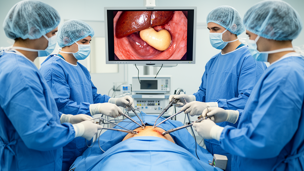
Management of the procedure required significant technical adjustment due to both the anatomical position and the presence of a large impacted stone in Hartmann’s pouch, which hindered traction and dissection. The surgical team employed a conventional four-port technique, which was augmented by a left-side-up table tilt to provide better visibility and access to the gallbladder. Meticulous dissection was conducted to ensure the strict identification of biliary structures. This disciplined approach was fundamental to the successful completion of the surgery.
The operative time for the procedure was 120 minutes, reflecting the technical challenges presented by the unexpected anatomy. Histopathological examination confirmed the presence of chronic cholecystitis, which was consistent with the patient’s reported history of recurrent biliary colic. Postoperatively, the patient exhibited an uneventful recovery. Follow-up examinations conducted three months after the surgery indicated that the patient remained well, with no signs of complications, confirming that the management strategies implemented were effective in addressing the surgical risks associated with this rare congenital anomaly.
The reported rate of bile duct injury during laparoscopic cholecystectomy in patients with a left-sided gallbladder has been noted as high as 4.4 percent, compared to the 0.3 to 0.5 percent risk associated with normally positioned gallbladders. Consequently, several intraoperative strategies have been established to enhance safety, including the use of a fundus-first approach, mirror-image port placement, and the liberal use of intraoperative cholangiography. These techniques are designed to minimize the possibility of damaging the common bile duct, which is susceptible to injury when the gallbladder is located in an atypical position.
Surgeons must maintain a high index of suspicion when encountering unusual anatomy during any laparoscopic cholecystectomy to prevent injury to the biliary tract. The case highlights that even in the absence of preoperative findings indicating an anomaly, intraoperative recognition is a critical skill for surgical teams. By applying meticulous dissection techniques and adhering strictly to the critical view of safety, surgeons can navigate the vascular and biliary variations inherent in true left-sided gallbladder cases. While the condition is a rare surprise during surgery, awareness of the associated embryological and anatomical possibilities provides the necessary foundation for ensuring successful and safe surgical outcomes.
In summary, the successful management of this case confirms that while true left-sided gallbladder in the medioposition is a rare biliary anomaly, it can be managed safely through standard laparoscopic approaches when paired with rigorous surgical caution. Despite the absence of preoperative evidence in most cases, the surgical team’s ability to adapt to unexpected findings demonstrates that the fundamentals of laparoscopic safety—specifically the critical view of safety—are essential for overcoming the elevated risks posed by these rare anatomical variations.
Beyond the primary complaint of recurrent abdominal pain, the preoperative clinical evaluation established important baseline findings for the patient. The authors noted that the forty-two-year-old Sri Lankan female had a documented three-year medical history of well-controlled diabetes mellitus alongside a body mass index of 36.5 kilograms per square meter. Despite experiencing recurrent biliary colic over a five-month span, her physical examination was unremarkable and routine liver function tests demonstrated completely normal values.
The published report details how early hepatic embryogenesis accounts for the structural variations frequently encountered during surgical exploration. Under ordinary developmental pathways, the gallbladder primordium originates from the hepatic diverticulum and migrates to occupy its standard position between liver segments IV and V. In true left-sided cases, the embryonic bud either migrates erroneously toward the left hepatic lobe or arises directly from the left ductal system. The researchers emphasize that this atypical migration directly explains accompanying anatomical variations documented across literature, which include anomalous cystic duct insertions, hypoplastic bile ducts, portal venous anomalies, and an unusual bile duct confluence located within the umbilical fissure.
During the operation, the team navigated complex vascular branching while securing structural orientation through standardized exposure methods. Alongside an impacted calculus in Hartmann’s pouch that hindered initial traction, operative findings demonstrated two distinct cystic arteries, comprising a short branch from the hepatic artery and an accessory vessel from a segment V arterial branch, in addition to a right hepatic artery crossing anterior to the common bile duct. To prevent ductal trauma, the surgical team documented the critical view of safety across two complementary angles: an anterior view delineating the cystic artery and a posterior view confirming the cystic duct, with the gallbladder situated on segment IVB to the right of the ligamentum teres and falciform ligament.
While alternative intraoperative strategies such as intraoperative cholangiography, mirror-image trocar placement, or a fundus-first approach are frequently cited to manage compromised visibility, this case confirmed that conventional four-port laparoscopy with left-side-up table positioning can achieve safe outcomes when surgeons strictly verify biliary anatomy.
Subject of Research: Case report of a rare true left-sided gallbladder in medioposition managed via laparoscopic cholecystectomy.
Article Title: True left-sided gallbladder (medioposition): a rare intraoperative biliary tract variation during laparoscopic cholecystectomy
Article References: Subasinghe, D., Wimalasena, G. H., & Maduwantha, P. (2026). True left-sided gallbladder (medioposition): a rare intraoperative biliary tract variation during laparoscopic cholecystectomy. Innovative Surgical Trends, 2(1), Article 9. https://doi.org/10.1007/s44414-026-00017-3
Image Credits: AI Generated
DOI: 10.1007/s44414-026-00017-3
Keywords: Cholecystectomy, Left-sided Gallbladder, Medioposition, Biliary Anomaly, Laparoscopic Surgery, Congenital Anomaly, Hepatic Anatomy, Vascular Variation, Critical View of Safety, Surgical Technique, Biliary Colic, Segment IVB, Ligamentum Teres, Case Report, Abdominal Surgery, Biliary Tract Variation, Chronic Cholecystitis
News Source: Ophelia Keating. (November 18, 2026). Surgeons Safely Manage Rare Left-Sided Gallbladder During Laparoscopic Cholecystectomy. Scienmag.