A cross-sectional clinical investigation involving 143 patients diagnosed with cerebral small vessel disease has demonstrated that integrating a five-minute virtual reality eye-tracking cognitive assessment with traditional magnetic resonance imaging burden scoring significantly improves the identification of mild cognitive impairment. Published in Frontiers in Human Neuroscience by a clinical research team from the Department of Neurology at Suzhou Hospital Affiliated to Anhui Medical University, the study evaluated whether combining objective functional measurements derived from visual exploration with established structural neuroimaging biomarkers could overcome the diagnostic limitations associated with evaluating imaging features alone. The investigators reported that while cerebral small vessel disease burden scoring alone yielded an area under the receiver operating characteristic curve of 0.874, combining the neuroimaging score with the composite virtual reality cognitive assessment score increased the area under the curve to 0.914, generating an apparent sensitivity of 87.1 percent and a specificity of 80.6 percent within the single-center cohort.
Cerebral small vessel disease encompasses a broad spectrum of pathological, clinical, and radiological processes that compromise the intracranial arterioles, capillaries, venules, and adjacent cerebral parenchyma. The condition represents the primary etiological driver of vascular cognitive impairment, which begins insidiously as mild cognitive impairment before potentially progressing toward vascular dementia. Pathophysiologically, the disorder is thought to originate from structural and functional disruption of the neurovascular unit, provoking chronic cerebral hypoperfusion, blood-brain barrier dysfunction, and low-grade neuroinflammation. These microvascular insults induce ischemic parenchymal injuries that manifest radiologically as white matter hyperintensities, lacunar infarcts, cerebral microbleeds, and enlarged perivascular spaces. Because vascular cognitive impairment is considered preventable and therapeutically controllable during its initial stages, establishing accurate and objective screening modalities capable of detecting early cognitive deficits in individuals with small vessel pathology is of urgent clinical significance.
Although magnetic resonance imaging remains the clinical cornerstone for identifying small vessel lesions in clinical practice, structural damage visualized on scans correlates imperfectly with clinical neurocognitive status. Prior investigations have demonstrated that individuals exhibiting comparable burdens of white matter hyperintensities or lacunar lesions can display divergent levels of cognitive functioning, largely due to variations in cognitive reserve, neural compensation, and underlying microstructural connectivity. In clinical settings, relying solely on total small vessel disease burden to detect mild cognitive impairment often suffers from suboptimal diagnostic specificity and sensitivity. In the present study, relying exclusively on the radiological burden score produced a specificity of only 66.7 percent, confirming that macrostructural imaging abnormalities cannot directly document actual cognitive performance or functional compensation, thereby underscoring the clinical necessity of establishing multimodal diagnostic frameworks that incorporate rapid, quantitative functional testing.
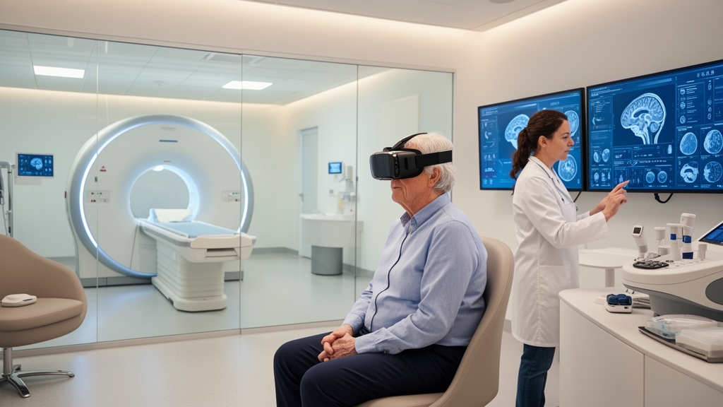
To capture functional cognitive deficits that elude structural imaging, the investigators implemented a computerized cognitive testing platform termed the virtual reality eye-tracking cognitive assessment, developed by Qingdao Zhisol Technology Company Limited. The apparatus integrates a Pico Neo 3 Pro Eye head-mounted display with embedded Tobii eye-tracking sensor technology to deliver an immersive, standardized testing environment that requires approximately five minutes to administer. During the testing paradigm, participants view three-dimensional simulated scenes containing target areas of interest alongside irrelevant distractors, with instructions directing them to identify and fixate upon the correct targets. The integrated eye-tracking module continuously registers gaze coordinates in real time, automatically calculating the proportion of time spent fixating on predefined target areas relative to overall gaze duration. By transforming objective ocular motor metrics into quantitative domain scores, the system captures dynamic executive processing and attentional allocation under high ecological validity.
The study protocol consecutively screened 149 patients presenting to the Department of Neurology at Suzhou Hospital Affiliated to Anhui Medical University between March 2025 and June 2026. All eligible participants were aged 50 years or older, had confirmed neuroimaging manifestations of cerebral small vessel disease on three-tesla cranial magnetic resonance imaging, possessed normal visual function, and were capable of cooperating with ocular movement tracking and scanning protocols. Individuals presenting with acute cerebral infarction lesions measuring 20 millimeters or greater on axial sequences, pre-existing dementia, or other neurological disorders such as Alzheimer’s disease, Parkinson’s disease, epilepsy, or multiple sclerosis were excluded. Six individuals were removed from the initial cohort because testing duration proved excessively prolonged, leaving 143 participants who completed the standardized clinical, radiological, and virtual reality evaluations.
Standard clinical classification divided the cohort into two distinct groups: a cognitively normal group consisting of 72 individuals and a mild cognitive impairment group containing 71 patients. Diagnostic categorization was established according to the 2018 Chinese Guidelines for the Diagnosis and Treatment of Dementia and Cognitive Impairment, executed independently by two trained neurologists blinded to the virtual reality eye-tracking results. Reference assessments incorporated the Beijing version of the Montreal Cognitive Assessment, the Chinese version of the Mini-Mental State Examination, the Clinical Dementia Rating scale, and the Activities of Daily Living scale. Patients allocated to the mild cognitive impairment group exhibited self-reported or informant-reported cognitive decline spanning more than three months, preserved basic daily functioning on the Activities of Daily Living scale with total scores under 26, a Clinical Dementia Rating of 0.5, and education-adjusted screening scores falling below established diagnostic cutoff thresholds.
Cranial neuroimaging was acquired on a three-tesla Philips Ingenia scanner using standardized sequence protocols including T1-weighted, T2-weighted, fluid-attenuated inversion recovery, diffusion-weighted, and susceptibility-weighted imaging. Scans were jointly analyzed by two blinded radiologists who reached diagnostic consensus. Total small vessel disease burden was scored on an ordinal four-point scale according to established Wardlaw criteria, awarding one point each for severe white matter hyperintensities graded by the Fazekas scale, the presence of one or more lacunar infarcts, the detection of cerebral microbleeds on susceptibility-weighted imaging, and moderate-to-severe enlarged perivascular spaces within the basal ganglia. In the analytical cohort, patients in the mild cognitive impairment group exhibited significantly higher median burden scores, increased Fazekas white matter damage, and higher prevalence rates of lacunar infarctions and cerebral microbleeds than those in the cognitively normal group.
Baseline demographic and clinical profiling revealed notable comparability between the two study groups, ensuring that diagnostic disparities were not attributable to conventional cardiovascular confounders. The cognitively normal group had a mean age of 68.44 years, while the mild cognitive impairment group had a mean age of 69.44 years, demonstrating no statistically significant difference. Similarly, distributions of biological sex, body mass index, years of formal education, and medical histories encompassing hypertension, diabetes mellitus, hyperlipidemia, coronary artery disease, prior ischemic stroke, tobacco smoking, and alcohol consumption showed no statistically significant differences between groups. In stark contrast, neuroimaging markers diverged markedly: patients with mild cognitive impairment demonstrated higher median total burden scores of three versus one, and higher rates of lacunar lesions at 70.42 percent versus 40.28 percent, reflecting greater underlying vascular structural damage.
Evaluation across the computerized functional battery demonstrated pronounced performance deficits among patients with mild cognitive impairment across all six evaluated cognitive domains. While traditional neuropsychological testing in small vessel disease often emphasizes psychomotor slowing, executive dysfunction, and attentional decline while sparing early episodic memory, the virtual reality assessment revealed widespread functional impairment. Overall mean virtual reality scores were significantly lower in the cognitively impaired group at 34.811 compared to 49.638 in the cognitively unimpaired group. Detailed domain evaluations showed statistically significant reductions in attention, abstract concept formation, mental calculation, executive control, short-term memory encoding, and delayed retrieval. The authors noted that navigating complex virtual scenes demands simultaneous recruitment of distributed brain networks, including the default mode and frontoparietal networks, rendering subtle microvascular network disruptions readily detectable.
To evaluate diagnostic utility, the investigators constructed a binary logistic regression model with total small vessel disease burden and overall virtual reality score entered as simultaneous independent predictors. The resulting omnibus test confirmed model significance, yielding a Nagelkerke pseudo-R-squared value of 0.631 and acceptable goodness-of-fit on the Hosmer-Lemeshow test. In multivariable analysis, higher virtual reality performance served as an independent protective factor against mild cognitive impairment classification, exhibiting an odds ratio of 0.932 per one-point increase. Conversely, each one-point increment in total small vessel disease burden score was associated with an elevated likelihood of impairment, demonstrating an odds ratio of 4.523. These findings confirmed that combining functional gaze metrics with structural imaging captures complementary pathological dimensions of small vessel disease.
Receiver operating characteristic curve analyses highlighted the incremental value of combining both testing modalities over individual parameters. When evaluated independently, the virtual reality score yielded an area under the curve of 0.779, with an optimal diagnostic threshold of 42.33 points or lower producing a sensitivity of 74.3 percent and a specificity of 70.8 percent. The total small vessel disease burden score achieved an area under the curve of 0.874, with a score threshold greater than one producing 88.7 percent sensitivity but a restricted specificity of 66.7 percent. When integrated into the multivariable logistic model, the combined diagnostic probability generated an area under the curve of 0.914. Pairwise DeLong tests demonstrated that this combined model significantly outperformed both the virtual reality score alone and the radiological burden score alone, achieving enhanced diagnostic balance without sacrificing case detection.
Despite these promising findings, the authors emphasized several methodological constraints inherent to the study design. Conducted as a single-center cross-sectional investigation without formal a priori sample size estimation, the sample of 143 participants may not fully reflect broader outpatient populations, and selection bias cannot be ruled out. Furthermore, because model parameters were derived and tested within the identical participant cohort without internal resampling or external cohort validation, the reported diagnostic statistics represent apparent performance estimates that require verification. The reference standard relied on education-adjusted screening scales rather than extensive neuropsychological testing batteries, and neuroimaging ratings were established by consensus without formal inter-rater reliability coefficients. Crucially, the cross-sectional architecture demonstrates statistical discrimination between pre-existing groups rather than prospective predictive accuracy, indicating that longitudinal multicenter studies are required to confirm whether virtual reality eye tracking can identify cognitive decline prior to standard clinical detection.
Subject of Research: Diagnostic efficacy of virtual reality eye-tracking assessment combined with total cerebral small vessel disease burden for mild cognitive impairment
Article Title: Application value of CSVD burden combined with virtual reality eye-tracking-based cognitive assessment in identifying mild cognitive impairment in patients with cerebral small vessel disease
Article References: Li, A., Wu, Q., & Yu, H. (2026). Application value of CSVD burden combined with virtual reality eye-tracking-based cognitive assessment in identifying mild cognitive impairment in patients with cerebral small vessel disease. Frontiers in Human Neuroscience, 20, Article 2002065. https://doi.org/10.3389/fnhum.2026.2002065
Image Credits: AI Generated
DOI: 10.3389/fnhum.2026.2002065
Keywords: cerebral small vessel disease, mild cognitive impairment, virtual reality eye tracking, CSVD burden, vascular cognitive impairment, magnetic resonance imaging, white matter hyperintensities, lacunar infarction, cerebral microbleeds, enlarged perivascular spaces, Tobii eye tracker, executive function, neurovascular unit, receiver operating characteristic, Pico Neo 3 Pro Eye, diagnostic modeling
News Source: Ophelia Keating. (November 18, 2026). Virtual Reality Eye Tracking and MRI Improve Mild Cognitive Impairment Identification. Scienmag.