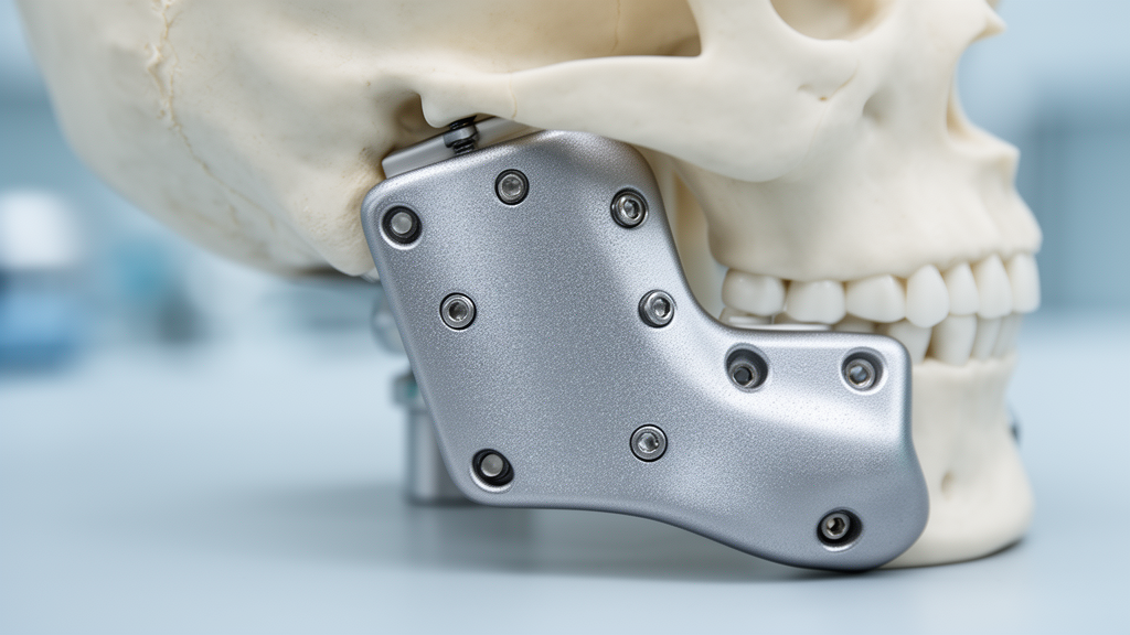
A prospective randomized clinical trial conducted at Alexandria University Teaching Hospital has demonstrated that customized, patient-specific three-dimensional titanium plates significantly reduce operative time and mitigate early postoperative morbidity in patients undergoing surgical repair for mandibular angle fractures. Published in BMC Oral Health, the clinical investigation compared sixteen adult patients allocated equally to receive either an individually milled three-dimensional titanium plate positioned according to a preoperative virtual surgical plan or a standard fixation construct consisting of two conventional titanium miniplates. While functional recovery, restored occlusion, and radiographic bone healing over four months remained equivalent between both treatment arms, patients receiving the personalized implants experienced an average surgical duration reduction of more than thirty-three minutes alongside measurably lower pain scores on the first postoperative day and reduced facial swelling across the initial postoperative week.
Mandibular fractures represent some of the most frequent traumatic injuries encountered across maxillofacial surgical units, second only to nasal bone fractures in overall incidence. Within this clinical category, fractures occurring at the mandibular angle account for approximately twenty-three to forty-two percent of all mandibular presentations, establishing them as particularly challenging clinical entities. The anatomical predisposition of the mandibular angle to mechanical failure arises from a convergence of structural factors, including a relatively reduced bony cross-sectional area, its functional role as the transition zone between the horizontal mandibular body and the vertical ramus, and the frequent impaction of third molars within the fracture line. When subjected to violent trauma—most commonly precipitated by road traffic collisions, interpersonal assaults, or accidental falls—these injuries disrupt the complex biomechanical equilibrium of the lower face, risking enduring deficits in masticatory capability, speech articulation, and normal swallowing if anatomical reduction and stable internal fixation are not successfully achieved.
The therapeutic paradigm for mandibular angle trauma has evolved from historic closed reduction methods and mandatory intermaxillary fixation toward open reduction and internal fixation utilizing osteosynthesis hardware. Standard surgical management frequently relies on miniplate fixation applied along the superior border following the principles established by Champy, which exploit natural physiological lines of tension. However, biomechanical investigations have highlighted that a solitary superior miniplate may provide inadequate rigidity against torsional stresses and dynamic multidirectional forces generated during jaw function, occasionally leading to complications such as hardware loosening, wound infection, malocclusion, or fibrous non-union. To mitigate these shortcomings, surgeons frequently employ a two-miniplate configuration that spans both the superior tension zone and the inferior compression zone. Nevertheless, adapting an inferior miniplate requires expanded soft-tissue exposure, transbuccal instrumentation using external trocar systems, or extraoral incisions, which elevate operative complexity and heighten surgical morbidity.
Recent breakthroughs in computer-aided design, computer-aided manufacturing, and additive or subtractive milling workflows have catalyzed the adoption of patient-specific fixation systems within craniomaxillofacial reconstruction. Rather than relying on standard prefabricated metal plates that necessitate laborious intraoperative manual bending and repetitive visual checks against exposed bone surfaces, computer-guided protocols harness three-dimensional patient anatomy derived from high-resolution computed tomography scans. This digital pipeline facilitates the fabrication of individualized titanium hardware engineered to conform precisely to the unique contours of the patient’s fractured bone. By incorporating virtual reduction planning, surgeons can predetermine the geometric configuration of the implant, optimize load distribution, and precalculate screw trajectories to prevent accidental injury to adjacent anatomical structures, including mandibular dental roots and the inferior alveolar neurovascular bundle traveling within the mandibular canal.
Conducted in accordance with updated CONSORT reporting recommendations and approved by the Research Ethics Committee of the Faculty of Dentistry at Alexandria University, this randomized trial enrolled sixteen non-medically compromised adult patients aged twenty to fifty years who presented between January 2025 and January 2026 with recent, non-infected, and non-comminuted mandibular angle fractures requiring open reduction and internal fixation. Patients presenting with pathological fractures, metabolic bone diseases, or severe systemic contraindications were excluded. Following informed written consent, participants were randomly allocated in a strict one-to-one ratio using computer-generated block randomization with block sizes of four. Allocation concealment was maintained through sequentially numbered, opaque, sealed envelopes. While the operating surgeons could not be masked to plate designs, independent clinical outcome evaluators and the biostatistician conducting the comparative data analyses remained fully blinded to treatment assignments.
The virtual workflow began with preoperative imaging performed on a sixteen-detector computed tomography scanner utilizing standardized tube voltage, current, and one-millimeter slice thicknesses with high-resolution bone reconstruction algorithms. Digital imaging files were imported into specialized segmentation software to generate three-dimensional virtual mandibular reconstructions, allowing investigators to achieve virtual anatomical reduction by confirming condylar seating within the glenoid fossa and restoring interarch dental relationships without interdental collision. Each custom plate was drafted with an integrated three-dimensional matrix featuring nine screw holes—five positioned along the superior margin and four along the inferior aspect—linked by rigid reinforcing struts. Milled from solid medical Grade IV titanium blocks by a specialized medical instrumentation manufacturer, the finished one-millimeter-thick implants were ultrasonically cleaned and autoclaved before surgery, completing an overall planning and production cycle of approximately forty-eight hours without delaying clinical care.
Surgical interventions for all sixteen participants were executed by the same principal maxillofacial surgeon to eliminate technical variance between cases. Both cohorts underwent general anesthesia with nasotracheal intubation and were approached via an intraoral vestibular incision along the external oblique ridge, followed by soft-tissue elevation and fracture segment mobilization. Following provisional maxillomandibular fixation to establish anatomical occlusion, patients assigned to the experimental group had their pre-sterilized custom plate seated along the superior tension zone and secured with predetermined monocortical screws. In contrast, patients in the control group received two conventional two-millimeter titanium miniplates: one manually contoured along the external oblique ridge and an inferior miniplate affixed along the basal border using a transbuccal trocar system. Associated contralateral fractures, present in select participants, were reduced and stabilized before angle fixation to restore dental arch continuity.
Both patient groups adhered to an identical, standardized postoperative care protocol to prevent pharmacological confounding. Therapeutic management encompassed a five-day regimen of systemic amoxicillin with clavulanic acid, metronidazole, alpha-chymotrypsin antiedematous therapy, and diclofenac potassium analgesia, supplemented by extraoral ice application during the first twenty-four hours, chlorhexidine antiseptic oral rinses, and a soft, high-calorie diet for four weeks. Clinical outcomes were comprehensively documented on postoperative days one, seven, fourteen, and thirty. Subjective pain severity was quantified utilizing a standard visual analogue scale ranging from zero to ten, while facial edema was tracked objectively using a calibrated linear tape measurement system summing three anatomical spans between the tragus, lateral ocular canthus, oral commissure, gonion angle, and soft-tissue pogonion against baseline values.
Analysis of surgical efficiency revealed a pronounced advantage for the digitally customized plate system. The mean intraoperative duration, measured meticulously from the start of fracture reduction to the final tightening of osteosynthesis screws, was 40.00 ± 9.35 minutes in the patient-specific cohort compared with 73.25 ± 11.22 minutes in the conventional two-miniplate cohort. This statistically significant difference of more than thirty-three minutes represented an approximate forty-five percent reduction in operative time. The experimental cohort also reported significantly less postoperative pain on day one, scoring 7.25 ± 0.71 versus 8.50 ± 0.93 in the control arm, with a mean difference of negative 1.25 points. Similarly, facial edema was significantly milder in the patient-specific group on postoperative day one (2.74 ± 0.40 centimeters versus 3.36 ± 0.46 centimeters) and day seven (1.31 ± 0.39 centimeters versus 1.70 ± 0.24 centimeters), though swelling differences between groups completely resolved by day fourteen.
Regarding the trial’s prespecified primary outcome, maximal mouth opening showed steady functional recovery in both cohorts, expanding from 20.38 ± 2.50 millimeters on postoperative day one to 39.63 ± 2.07 millimeters on day thirty in the patient-specific group, and from 19.50 ± 2.78 millimeters to 39.13 ± 2.90 millimeters in the conventional miniplate group. Repeated-measures analysis of variance confirmed a powerful effect of time but no significant overall difference between treatment groups or group-by-time interaction. Postoperative dental occlusion was successfully restored and maintained in all sixteen participants without discrepancies such as premature contacts or anterior open bites. Soft-tissue healing evaluated via the Landry Wound Healing Index also progressed similarly across cohorts. One patient in each study arm developed localized superficial wound dehiscence during the first postoperative week, and both instances resolved uneventfully under conservative local wound debridement, saline irrigation, and oral hygiene reinforcement.
Quantitative radiographic assessments affirmed that biomechanical stability was fully preserved with the customized single-plate design. Baseline computed tomography scans acquired immediately after operation verified accurate anatomical reduction, while four-month follow-up scans demonstrated continuous osseous bridging across all fracture sites without any instances of non-union, malunion, or hardware displacement. Bone mineral density, quantified objectively in Hounsfield units at three standardized regions of interest along the superior, middle, and inferior thirds of the fracture line while avoiding metal-induced scatter artifacts, exhibited robust osteogenic healing. Mean bone density rose from 661.1 ± 68.24 to 1075.1 ± 111.7 Hounsfield units in the patient-specific group and from 656.0 ± 62.10 to 1100.3 ± 92.27 Hounsfield units in the two-miniplate group, demonstrating statistically equivalent mineralization trajectories across both cohorts.
The authors acknowledged several key methodological limitations inherent to the trial. The modest sample size of sixteen patients treated at a single university center limits statistical power for subtle secondary endpoint variations and restricts broader geographic generalizability. Furthermore, while the four-month follow-up window proved sufficient to establish primary union and early soft-tissue resolution, it remained inadequate to detect delayed hardware complications, screw loosening, or long-term fatigue performance under masticatory forces. The study also noted the retrospective timing of its trial registry entry on ClinicalTrials.gov and emphasized that computed tomography density values serve as surrogate markers of radiographic mineralization rather than absolute biological or mechanical proof of cortical maturity. Additionally, formal health economic calculations were absent; future multicenter investigations must therefore assess whether operating room time savings offset the specialized software licenses and custom manufacturing expenditures required for personalized craniomaxillofacial hardware.
Subject of Research: Comparison of patient-specific three-dimensional titanium plates and conventional two-miniplate fixation for mandibular angle fractures
Article Title: Patient-specific three-dimensional titanium plate versus conventional two-miniplate fixation for mandibular angle fractures: a randomized clinical trial
Article References: Abd Elfattah, A. M., Eldibany, M. M., Hassan, R. S., & Oraby, M. S. (2026). Patient-specific three-dimensional titanium plate versus conventional two-miniplate fixation for mandibular angle fractures: a randomized clinical trial. BMC Oral Health, 26(1), Article 2291. https://doi.org/10.1186/s12903-026-10155-2
Image Credits: AI Generated
DOI: 10.1186/s12903-026-10155-2
Keywords: mandibular angle fractures, patient-specific implants, three-dimensional titanium plates, two-miniplate fixation, open reduction internal fixation, randomized clinical trial, maxillofacial surgery, computer-aided design, virtual surgical planning, intraoperative time, maximal mouth opening, facial edema, bone density, Hounsfield units, Champy principles, transbuccal trocar, wound healing
News Source: Ophelia Keating. (November 18, 2026). Custom 3D Titanium Plates Accelerate Mandibular Angle Fracture Surgery in Trial. Scienmag.