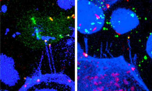
During embryonal development of vertebrates, signaling molecules inform each cell at which position it is located. In this way, the cell can develop its special structure and function. For the first time now, researchers of Karlsruhe Institute of Technology (KIT) have shown that these signaling molecules are transmitted in bundles via long filamentary cell projections. Studies of zebrafish of the scientists of the European Zebrafish Resource Center (EZRC) of KIT revealed how the transport of the signaling molecules influences signaling properties. A publication in the Nature Communications journal presents the results.
Control of cell differentiation in the central nervous system. Photo Credit: Image courtesy of Karlsruhe Institute of Technology
Organisms, organs, and tissues are complex three-dimensional systems that consist of thousands of cells of various types. During embryonal development of vertebrates, each cell requires information on the position at which it is located in the tissue. This position information enables the cell to develop a certain cell type for later execution of the correct function. This information is transmitted via signal molecules, so-called morphogenes. These morphogenes are not homogenously distributed in the tissue, their concentration varies. Various concentrations activate various genes in the target cell.
The cells in the developing central nervous system receive their position information from signal molecules belonging to the family of Wnt proteins. The concentration of Wnt proteins determines whether a cell differentiates to a cell of the forebrain or of the afterbrain. “Distribution of these signal molecules has to be controlled precisely,” Dr. Steffen Scholpp, head of a research group of the KIT Institute of Toxicology and Genetics (ITG), explains. “Smallest changes of the concentration or the transport direction may cause severe damage, such as massive malformations during embryonal development or formation of cancer.”
For the first time now, the working group of Dr. Steffen Scholpp has shown that the Wnt proteins are transmitted specifically via long cell projections, so-called filopodia. In the Nature Communications journal, the scientists report that the signaling factors are loaded on the tips of the filopodia only. In this way, signaling can start immediately upon contacting. The signaling factors bind to the corresponding receptors of the target cell and induce the correct cell response. “Now, the source cell can decide precisely which target cell receives how much signaling protein at which time,” Scholpp explains. The KIT researchers study zebrafish and human cell lines and succeeded in reproducing or reducing the filopodia and analyzing the resulting changes of signaling properties of the Wnt morphogenes.
Story Source:
The above story is based on materials provided by Karlsruhe Institute of Technology.


