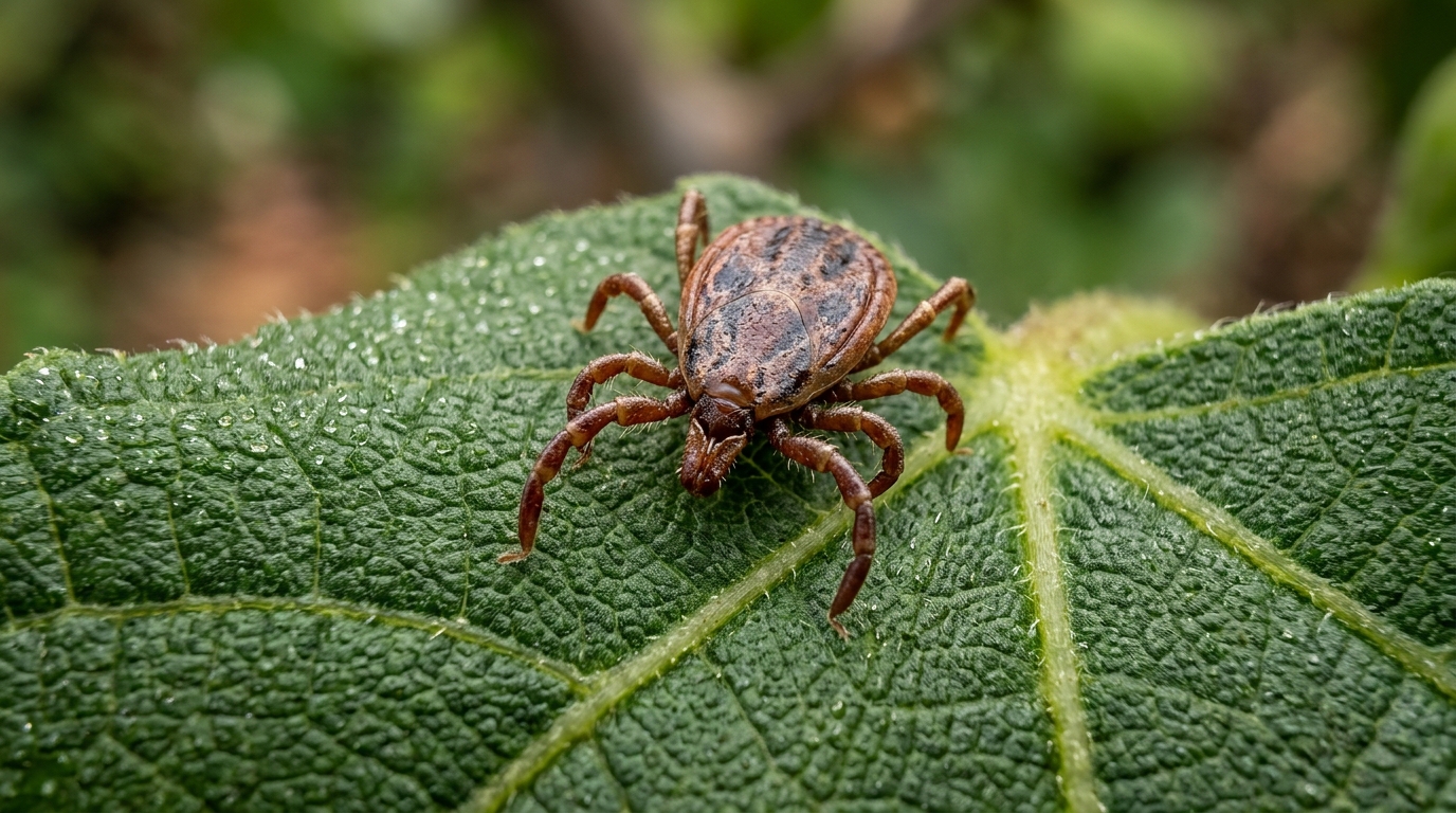
A newly published laboratory investigation demonstrates that silver nanoparticles synthesized using an aqueous leaf extract from the common fig tree achieve complete mortality against larvae of the cattle tick Rhipicephalus microplus while substantially inhibiting egg laying in adult females. Conducted by an international team of researchers, the research evaluated the acaricidal efficacy of biologically engineered nanomaterials alongside computational evaluations of phytochemical binding against an essential tick receptor protein. The study investigated the physical and chemical properties of the green-synthesized particles and assessed their biological performance across multiple concentrations under controlled environmental conditions. By pairing standard immersion and packet bioassays with molecular docking and pharmacokinetic screening, the investigation established both the lethal capacity of the nanoparticle formulations and the specific organic compounds responsible for particle stabilization.
The cattle tick Rhipicephalus microplus represents one of the most economically damaging ectoparasites affecting cattle across tropical and subtropical regions. Infestations lead to severe production losses in domestic herds by reducing weight gain, decreasing overall milk yields, and causing anemia through direct blood loss. However, extensive chemical interventions have triggered widespread physiological resistance among tick populations worldwide, while generating persistent toxic residues in dairy products, commercial meat, and local ecosystems.
To generate the nanostructures, researchers collected fresh botanical specimens of Ficus carica from District Mardan within Pakistan, depositing an identified reference voucher at Abdul Wali Khan University Mardan under accession Awkum Bot 171 1 11. The aqueous extraction procedure involved washing, chopping, and boiling ten grams of clean foliage in one hundred milliliters of double-distilled water for five minutes before filtration through standard Whatman filter paper. Synthesis proceeded by mixing the clear botanical filtrate with a one millimolar solution of silver nitrate at a specific one-to-two-hundred ratio, maintaining the mixture at a stable pH of 6.8 under ambient room lighting at twenty-five degrees Celsius. Over twenty-four hours of incubation, the reaction mixture transitioned visually from a light yellow liquid into an intense dark brown colloidal suspension. This distinct optical transition served as primary macroscopic evidence confirming the reduction of silver cations into elemental metallic silver nanoparticles, which were subsequently harvested through high-speed centrifugation, washed, and dried.
Spectroscopic characterization confirmed the formation, optical properties, and surface chemistry of the synthesized metallic nanomaterials. Ultraviolet-visible absorption spectroscopy performed across wavelengths from two hundred to six hundred nanometers revealed a prominent surface plasmon resonance peak centered at four hundred twenty-nine nanometers. The intensity of this absorption band steadily increased during the twenty-four-hour reaction timeframe, confirming continuous particle nucleation and kinetic equilibrium without altering the fundamental absorbance profile. Fourier-transform infrared spectroscopy documented several major vibrational bands reflecting the presence of organic functional groups derived from the plant broth. Prominent bands appeared at 3349.7 inverse centimeters representing hydroxyl group stretching vibrations, 1750.6 inverse centimeters corresponding to carbonyl bond stretching, and 1279.5 and 1036.6 inverse centimeters associated with carbon-oxygen and carbon-nitrogen linkages. A fingerprint band at 663.9 inverse centimeters indicated organic bending modes, confirming that native phytochemicals including polyphenols and organic acids successfully capped and stabilized the inorganic silver cores against excessive aggregation.
Structural and morphological evaluations provided detailed measurements of the crystalline organization and physical dimensions of the manufactured nanoparticles. X-ray diffraction patterns displayed five distinct Bragg reflection peaks at two-theta angles of 38.1, 44.3, 64.5, 77.5, and 81.7 degrees, corresponding precisely to the 111, 200, 220, 311, and 222 lattice planes of face-centered cubic metallic silver recorded under standard crystallographic databases. Calculating peak broadening via the Debye-Scherrer equation established an average crystallite domain size ranging between twenty-one and twenty-six nanometers. Morphological imaging conducted through scanning electron microscopy showed predominantly spherical particles exhibiting mild clustering, with individual nanostructures spanning thirty to eighty-five nanometers and displaying an average polydisperse diameter of five hundred nanometers with a standard deviation of 355.9 nanometers. Complementary energy-dispersive X-ray spectroscopy identified a characteristic silver emission peak at three kiloelectronvolts alongside secondary carbon signals attributable to the organic capping layer and sample substrate.
Chemical profiling of the botanical leaf extract utilizing gas chromatography-mass spectrometry clarified the specific bioactive molecules responsible for metal ion reduction and particle capping. The chromatographic evaluation separated eight prominent elution peaks that permitted the structural identification of twelve distinct phytocompounds compared against authenticated spectral libraries. Key identified constituents included 1,2-benzenediol, 2H-1-benzopyran-2-one, hexanedioic acid, acridine, benzeneethanamine, metaraminol, p-hydroxyamphetamine, 1-isopropyldiaziridine, 1-methyl-2-phenoxyethylamine, and 2,8,9-trioxa-5-aza-1-silabicyclo. Mechanistically, electron-dense hydroxyl moieties located on 1,2-benzenediol facilitate direct electron transfer to convert silver ions into elemental atoms through oxidation into quinone intermediates. Concurrently, dicarboxylic structures such as hexanedioic acid coordinate silver ions across oxygen atoms, while amine-bearing molecules like metaraminol and heterocyclic structures like coumarin donate electron density from nitrogen and carbonyl groups, stabilizing newly nucleated crystalline surfaces and preventing excessive particulate agglomeration during ambient synthesis.
The biological potency of the green-synthesized silver nanoparticles was initially quantified through standardized larval packet bioassays using fourteen-to-twenty-one-day-old Rhipicephalus microplus larvae hatched from laboratory-incubated eggs. Treated Whatman filter paper packets were impregnated with suspension concentrations ranging from five to sixty milligrams per milliliter, with controls receiving distilled water or the synthetic pyrethroid cypermethrin. Following twenty-four hours of continuous packet exposure within a controlled environmental chamber maintained at twenty-eight degrees Celsius and eighty percent relative humidity, the experimental formulations demonstrated strong, concentration-dependent larvicidal action. The highest applied concentration of sixty milligrams per milliliter induced complete one-hundred-percent larval mortality, successfully matching the lethal performance exhibited by commercial cypermethrin. Probit regression analysis confirmed that larval death rates climbed consistently with increasing exposure levels, demonstrating that the botanical nanoparticle complex reliably disrupted larval survival under closed packet conditions without requiring synthetic chemical adjuvants.
Adult immersion tests conducted on fully engorged adult female ticks gathered from field cattle demonstrated substantial reproductive impairment following direct short-term contact with the nanoparticle suspensions. Adult females immersed for five minutes in concentrations between five and forty milligrams per milliliter exhibited marked reductions in subsequent oviposition activity compared to untreated controls. When administered at the maximum immersion dosage of forty milligrams per milliliter, the botanical silver nanoparticle treatment achieved an oviposition inhibition rate of 84.79 percent, closely approaching the 87.535 percent inhibition observed in cypermethrin-treated positive controls. Furthermore, the mean egg weight index dropped to 0.198 grams among treated females, compared to 0.394 grams in the distilled water control cohort, representing a statistically significant reproductive reduction at a p-value below 0.05. These quantitative outcomes demonstrate that direct topical exposure to the nanoparticle suspension severely compromises female egg-laying capability and overall reproductive viability.
To explore the molecular mechanisms underlying the observed tick mortality and reproductive failure, researchers conducted in silico homology modeling and automated ligand docking against the tick Membrane Associated Progesterone Receptor protein. A three-dimensional structural model generated from the accession XP 037276930.1 sequence achieved an ERRAT overall quality factor of 95.918, with Ramachandran plot validation confirming 87.2 percent of amino acid residues in favored regions and zero residues in disallowed space. CASTp pocket searches mapped critical catalytic pocket residues, including tyrosine 116, valine 133, and lysine 164. Docking simulations performed in AutoDock Vina revealed that the plant coumarin 2H-1-benzopyran-2-one bound strongly within this active site, yielding an affinity of minus 8.2 kilocalories per mole stabilized by hydrogen bonding with tyrosine 164 and hydrophobic pi stacking. Notably, this botanical ligand exhibited higher binding affinity than cypermethrin, which registered minus 7.9 kilocalories per mole.
In silico pharmacokinetic screening using the Deep-PK platform indicated that the majority of identified phytocompounds adhered to Lipinski rules of five for drug-likeness, maintaining favorable molecular weights between 86.13 and 270.39 grams per mole and appropriate lipophilicity with calculated LogP values spanning 0.029 to 4.88. Compounds such as 1,2-benzenediol and hexanedioic acid displayed favorable predictive safety profiles regarding mutagenicity and hepatotoxicity, though every compound exhibited potential dermal sensitization, and acridine showed flags for mutagenic risk. Importantly, the authors emphasize that while these in vitro and computational data demonstrate notable acaricidal efficacy, the study remains constrained by the absence of live animal trials, mammalian toxicity evaluations, and long-term ecological assessments. Because dispersed silver nanoparticles pose recognized environmental hazards to beneficial aquatic organisms and soil microbial communities, comprehensive field trials and environmental safety investigations remain mandatory before these botanical nanomaterials can be deployed in livestock management.
Subject of Research: Biosynthesis and acaricidal evaluation of Ficus carica silver nanoparticles against cattle ticks
Article Title: Biosynthesis, characterization, and acaricidal activity of silver nanoparticles from Ficus carica leaf extract against Rhipicephalus (Boophilus) microplus ticks, a Vector of Bacterial Pathogens”
Article References: Malak, N., Ullah, S., Qarni, A., Khan, A., Niaz, S., Bayugar, R. C., Ali, H., Said, M. A., Alqahtani, F. M., Younous, Y. A., & Khan, A. (2026). Biosynthesis, characterization, and acaricidal activity of silver nanoparticles from Ficus carica leaf extract against Rhipicephalus (Boophilus) microplus ticks, a Vector of Bacterial Pathogens”. Discover Nano, 21(1), Article 577. https://doi.org/10.1186/s11671-026-05049-9
Image Credits: AI Generated
DOI: 10.1186/s11671-026-05049-9
Keywords: Ficus carica, silver nanoparticles, Rhipicephalus microplus, green nanotechnology, acaricidal activity, larval packet bioassay, adult immersion test, oviposition inhibition, molecular docking, RmMAPRC, 2H-1-benzopyran-2-one, cypermethrin, cattle tick control, surface plasmon resonance, phytochemical reduction, Deep-PK screening
News Source: William Thompson. (November 16, 2026). Fig Leaf Silver Nanoparticles Eliminate Cattle Ticks in Laboratory Bioassays. Scienmag.