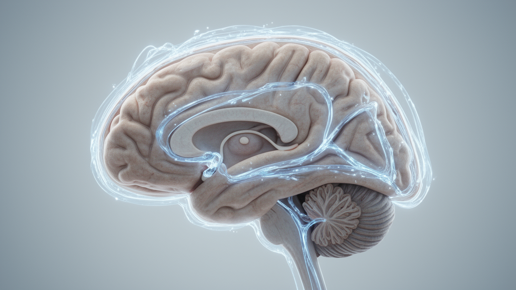
In a human neuroimaging study published in Nature Aging, researchers demonstrated that the rhythmic waves of cerebrospinal fluid flowing through the brain during sleep decline substantially with age, occurring before the onset of diagnosed neurological conditions. By recording synchronized electrical activity and accelerated hemodynamic signals across younger and older cohorts, the study identified a dual impairment spanning neural oscillations and cerebrovascular dynamics that together diminish fluid movement during non-rapid eye movement sleep. In healthy young individuals, large-amplitude fluid pulses surge through the ventricular system during non-rapid eye movement sleep, but the researchers found that these waves become markedly blunted in the aging brain. The investigation establishes that the degradation of sleep-related brain fluid dynamics is not an incidental marker of neurodegenerative pathology, but an early physiological transformation emerging in typical human aging.
The physiological process propelling fluid movement through the brain involves a tightly coordinated neurovascular cascade operating predominantly during deep, non-rapid eye movement rest. Synchronized slow-wave neural activity prompts rhythmic contractions and dilations within cerebral blood vessels, generating large-scale shifts in cerebral blood volume through local neurovascular coupling, autonomic influences, and neuromodulatory rhythms. When cerebral blood volume decreases, cerebrospinal fluid rushes inward to compensate, whereas vascular expansion drives fluid outward. Although prior studies established that healthy young adults routinely demonstrate prominent low-frequency cerebrospinal fluid waves at approximately 0.05 hertz during non-rapid eye movement sleep, scientific understanding remained incomplete regarding whether healthy aging alters this sleep-dependent circulatory mechanism in the absence of diagnosed disease.
To address this question, an investigative team led by Sydney M. Bailes, Stephanie D. Williams, and Laura D. Lewis collected simultaneous electroencephalography and fast functional magnetic resonance imaging data during nighttime sleep across two research sites. Both sites, Boston University and the Massachusetts Institute of Technology, utilized identical 3-Tesla Siemens Prisma scanners equipped with 64-channel head coils, matched functional sequences, and synchronized 32-channel MR-compatible electroencephalography caps. Scans began around midnight, and participants avoided naps and caffeine consumption after 11:00 on experiment days. After applying exclusion criteria for motion artifacts and persistent wakefulness, the analyzed nighttime cohort comprised thirty-one young adults between 18 and 39 years of age, with a mean age of 24 years, and forty-one older adults between 60 and 83 years of age, with a mean age of 69 years. Statistical testing confirmed that the primary outcome measures were robust across scanner locations, showing no significant differences attributable to the testing site.
To track cerebrospinal fluid movement non-invasively, the researchers implemented an accelerated gradient-echo echo-planar imaging sequence featuring a repetition time of 0.378 seconds, an echo time of 31 milliseconds, 2.5-millimeter isotropic voxels, and a multiband acceleration factor of eight. This protocol leverages flow-related enhancement, an imaging effect wherein rapidly moving fluid entering the edge slice of an imaging volume appears bright because it has not experienced saturation from repetitive radiofrequency pulses. Positioning the bottom slice of the imaging volume to intersect the fourth ventricle enabled sensitive detection of dorsal cerebrospinal fluid inflow while simultaneously recording blood-oxygen-level-dependent hemodynamic signals across cerebral gray matter. Ventricular regions of interest were delineated manually on single-band reference scans lacking multiband acceleration, avoiding selection bias based on time-series information. These manual masks were registered to automated anatomical segmentations using boundary-based and non-linear registration tools in native participant space, ensuring that fluid inflow signals and cortical hemodynamics were extracted with high spatial fidelity.
Spectral power analysis across continuous 60-second intervals of wakefulness and non-rapid eye movement sleep revealed pronounced differences in fluid dynamics between cohorts. Multitaper spectral estimation demonstrated that in young adults, low-frequency cerebrospinal fluid power between 0.01 and 0.1 hertz increased significantly by an average of 3.82 decibels upon entering non-rapid eye movement sleep. In striking contrast, older adults exhibited an average change of only 1.15 decibels across the transition from wakefulness to sleep, representing a non-significant state change. A linear mixed-effects model revealed a significant age-by-state interaction, demonstrating that the age-related reduction in cerebrospinal fluid flow was significantly stronger during sleep than during wakefulness. Furthermore, older participants had significantly lower fluid power during non-rapid eye movement sleep than young adults. A secondary control analysis dividing fluid signals by ventricular cross-sectional area confirmed that anatomical differences in fourth ventricle dimensions did not explain this marked functional reduction.
Because cerebral blood volume fluctuations mechanically propel ventricular fluid, the research team examined whether cortical hemodynamic waves exhibited parallel age-related reductions during sleep. In young adults, low-frequency blood-oxygen-level-dependent power within gray matter increased by an average of 3.67 decibels from wakefulness to sleep, whereas older adults achieved a significantly smaller sleep-related increase of 2.15 decibels. Evaluating temporal dynamics across stable 90-second intervals of non-rapid eye movement sleep demonstrated that young adults displayed tight cross-correlations between ventricular cerebrospinal fluid inflow and the zero-thresholded negative derivative of the gray matter hemodynamic signal, achieving a maximal correlation coefficient of 0.67 at a lag of negative 1.13 seconds. In older adults, this cross-correlation reached only 0.42 at a lag of negative 0.76 seconds, representing a statistically significant attenuation of coupling strength. Even after matching motion profiles between cohorts through nearest-neighbor resampling of framewise displacement, this mechanical decoupling between gray matter vascular oscillations and ventricular fluid pulsations persisted in older individuals.
Parallel electrophysiological measurements identified significant degradation in the electrical slow waves that normally initiate this neurovascular cascade. Frontal electroencephalography slow-wave power within the 0.5 to 4 hertz band, computed across seven frontal electrodes including F3, F4, F7, F8, Fz, FT9, and FT10, increased during non-rapid eye movement sleep in both groups but was substantially lower in older adults. Cross-correlation between the envelope of frontal slow-wave activity and subsequent cerebrospinal fluid oscillations revealed that electrical slow waves reliably preceded fluid motion in both cohorts, confirming the directional hierarchy of the neurovascular pathway. However, the maximal correlation between frontal neural slow waves and fluid pulsations fell from 0.23 at a lag of 8.69 seconds in young adults to just 0.09 at a lag of 9.45 seconds in older adults. In both young and older participants, individual frontal slow-wave power correlated positively with the magnitude of low-frequency fluid flow, reinforcing the direct dependency of fluid propulsion on synchronized cortical slow waves.
To determine whether fluid impairment stemmed directly from neural slow-wave loss or vascular dysfunction, the researchers constructed a serial mediation model using structural equation modeling. The model evaluated three concurrent pathways: an independent neural pathway, an independent hemodynamic pathway, and a serial neurovascular sequence linking age, electroencephalography slow-wave power, gray matter hemodynamics, and ventricular fluid flow. The analysis revealed that age exerted a significant indirect effect on cerebrospinal fluid flow transmitted serially through the neurovascular pathway, wherein age-related loss of slow-wave power led to attenuated hemodynamic oscillations that subsequently reduced fluid movement. Furthermore, frontal slow-wave power significantly mediated the age-related reduction in fluid flow independently of hemodynamics. Conversely, gray matter hemodynamics did not mediate fluid loss once slow-wave power was accounted for, indicating that sleep-dependent hemodynamic decline occurs as a secondary consequence of diminished neural driving forces. A statistically significant direct effect of age on fluid flow persisted, suggesting that additional biological mechanisms contribute to fluid decline.
To establish whether neurovascular responses were causally impaired independently of spontaneous sleep states, the investigators conducted daytime waking experiments on thirty-one young and fifty-five older participants using controlled sensory stimulation. Participants viewed an inverting checkerboard stimulus flickering at 12 hertz across alternating 16-second active blocks and 30-to-32.5-second resting periods. Analysis of the stimulus-evoked neural envelope across occipital electroencephalography channels demonstrated equivalent electrical responses between younger and older adults, confirming that sensory cortical excitation was matched. Despite identical neural activation, older adults exhibited significantly smaller hemodynamic responses in the primary visual cortex and significantly reduced task-evoked cerebrospinal fluid inflow in the fourth ventricle. At the individual participant level, the magnitude of sensory-evoked fluid flow during wakefulness correlated significantly with low-frequency fluid power during nighttime non-rapid eye movement sleep within the older cohort. This daytime experiment proved that impaired neurovascular coupling directly restricts fluid movement in the aging brain even when neural driving inputs remain intact.
In an accompanying daytime challenge, the research team evaluated intrinsic cerebrovascular reactivity using an end-inspiration breath-holding task consisting of eight 15-second breath-holds while continuously monitoring expired carbon dioxide. Quantitative mapping revealed that gray matter cerebrovascular reactivity was significantly reduced in older adults compared with young adults, accompanied by blunted post-breath-hold hemodynamic and ventricular fluid responses. Structural analyses further demonstrated that normalized frontal gray matter volume was significantly lower in older adults and correlated selectively with nighttime fluid flow, whereas other lobes showed no relationship. Despite group-level reductions, substantial individual variability existed, with some older adults exhibiting youthful slow-wave power or intact vascular reactivity. The authors noted that while macroscopic ventricular flow was measured, microscopic waste clearance was not quantified, and variables like blood pressure warrant future study. Distinct mechanistic origins indicate that interventions targeting brain fluid physiology must be tailored to specific neural or vascular deficits.
Subject of Research: Age-related decline in sleep-dependent cerebrospinal fluid flow and hemodynamics
Article Title: The aging human brain exhibits reduced hemodynamic and cerebrospinal fluid flow waves during sleep
Article References: Bailes, S. M., Williams, S. D., Ashenagar, B., Pham, T. L. D., Valdiviezo, Z., Licata, J., Bosli, M. Y., Dormes, B. J., Yun, H. J., Zimmerman, D., Hernandez Moyers, A., Salat, D. H., & Lewis, L. D. (2026). The aging human brain exhibits reduced hemodynamic and cerebrospinal fluid flow waves during sleep. Nature Aging. https://doi.org/10.1038/s43587-026-01270-4
Image Credits: AI Generated
DOI: 10.1038/s43587-026-01270-4
Keywords: cerebrospinal fluid, sleep physiology, hemodynamic oscillations, brain aging, slow-wave sleep, non-rapid eye movement, fourth ventricle, blood-oxygen-level-dependent, cerebrovascular reactivity, neurovascular coupling, electroencephalography, functional magnetic resonance imaging, frontal cortex, gray matter atrophy, fluid dynamics, neural oscillations, breath-hold task
News Source: Cassandra Pierce. (November 16, 2026). Aging Brain Shows Reduced Cerebrospinal Fluid Waves During Nighttime Sleep. Scienmag.