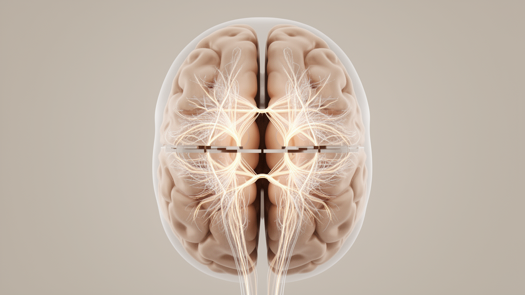
In a resting-state functional neuroimaging investigation into persistent postural-perceptual dizziness, patients exhibited significantly reduced functional cooperation between corresponding regions across the two cerebral hemispheres, particularly within visual and multisensory cortical territories. The investigation, published in Frontiers in Neurology, evaluated macroscale interhemispheric coordination and functional lateralization in forty-seven patients with persistent postural-perceptual dizziness and fifty-two matched healthy controls. Using an advanced computational framework termed connectivity between functionally homotopic voxels, researchers identified marked functional decoupling between contralateral homologous areas in visual and associative parietal structures. However, the study authors emphasize an essential interpretive qualification: these interhemispheric connectivity differences did not survive cluster-level family-wise error correction when statistical models were adjusted for comorbid anxiety, depressive symptoms, or subjective sleep disturbance. Consequently, while the study establishes a distinct pattern of disrupted bilateral sensory synchronization in unadjusted analyses, the findings highlight an unresolved question regarding whether these alterations reflect core dizziness pathophysiology or secondary psychological disruptions.
Persistent postural-perceptual dizziness, formally recognized as a chronic functional vestibular disorder by the Bárány Society in 2017, represents a common cause of persistent, non-spinning vertigo and unsteadiness encountered in neurological clinics. Patients endure chronic dizziness, unsteadiness, or floating sensations lasting at least three months, which are typically exacerbated by upright posture, active or passive movement, and exposure to complex or moving visual environments. Despite marked impairment in daily functioning, postural stability, and spatial orientation, standard clinical examinations and routine peripheral vestibular tests reveal no overt structural brain lesions or acute end-organ damage. Contemporary neuroscientific models propose that the condition involves maladaptive neuroplastic changes following an initial vestibular or psychological event, resulting in abnormal sensory weighting, visual dependence, and faulty integration across vestibular, visual, and somatosensory networks. While prior functional neuroimaging studies have shown altered regional network activity, higher-order organizational features such as hemispheric specialization and interhemispheric cooperation have remained poorly characterized.
To investigate these organizational dynamics, researchers recruited fifty-four adult patients meeting the 2017 diagnostic criteria from the specialized dizziness clinic at the First Affiliated Hospital of the University of Science and Technology of China between January 2023 and December 2025. Following clinical exclusions and imaging quality control, forty-seven patients with persistent postural-perceptual dizziness and fifty-two healthy controls were included in the final analysis. The two cohorts were closely matched in chronological age, sex distribution, and educational attainment. Clinical assessments revealed substantial differences between the groups: patients exhibited significantly higher scores on the 14-item Hamilton Anxiety Rating Scale, the 24-item Hamilton Depression Rating Scale, the Pittsburgh Sleep Quality Index, and the Dizziness Handicap Inventory. Cognitive evaluations showed modestly lower scores on the Beijing version of the Montreal Cognitive Assessment among patients, whereas scores on the Mini-Mental State Examination showed no statistically significant group differences.
Neuroimaging acquisition was performed using a 3.0-Tesla Discovery MR750w magnetic resonance imaging scanner equipped with a twenty-four-channel head coil. High-resolution anatomical scans were acquired via three-dimensional fast spoiled gradient-recalled echo sequences to enable structural spatial normalization and tissue segmentation, while blood-oxygen-level-dependent resting-state functional volumes were collected using single-shot echo-planar imaging sequences across an eight-minute scan duration. Image preprocessing was carried out using the WhiteMatter toolkit, integrating procedures from SPM12, AFNI, and FSL. The preprocessing pipeline included removing the first five functional volumes, despiking, slice-timing correction, motion realignment, and coregistration of functional images to structural volumes. The researchers regressed out twenty-seven nuisance regressors, including signals from white matter, cerebrospinal fluid, global brain volume, and the Friston twenty-four-parameter motion model, followed by Gaussian spatial smoothing, band-pass filtering, and spatial normalization into Montreal Neurological Institute coordinate space. Motion analyses verified that no included participant exceeded translation thresholds of three millimeters or three degrees.
The study evaluated two complementary neuroimaging metrics to capture bilateral functional organization across the cerebral cortex: the autonomy index and connectivity between functionally homotopic voxels. The autonomy index measures the relative predominance of functional connections within the same hemisphere compared to connections reaching into the contralateral hemisphere, with higher values indicating greater regional functional autonomy and hemispheric specialization. In contrast, connectivity between functionally homotopic voxels quantifies functional coupling between functionally corresponding anatomical locations across the two hemispheres. Traditional techniques such as voxel-mirrored homotopic connectivity assume strict geometric symmetry by pairing coordinates across an anatomically mirrored mid-sagittal plane, which can overlook individual variations in cortical folding and functional topography. By identifying functional homologues based directly on empiric functional connectivity profiles derived across the entire pooled sample, connectivity between functionally homotopic voxels evaluates synchronized activity between true functional counterparts regardless of minor structural asymmetries.
In primary whole-brain analyses adjusting for participant age, sex, and mean framewise displacement, voxel-wise nonparametric permutation tests revealed significant reductions in connectivity between functionally homotopic voxels among patients compared to healthy controls. These reductions in interhemispheric coordination appeared across six distinct anatomical clusters, including the right calcarine cortex, the right cuneus, the left inferior occipital gyrus, the left middle occipital gyrus, the left inferior parietal lobule, and the right middle temporal gyrus. Statistical differences in all six clusters remained significant under family-wise error correction at a cluster-level threshold below 0.05. These regions represent critical nodes supporting primary visual processing, visuospatial attention, and the integration of visual, vestibular, and somatosensory inputs. In contrast, when the researchers analyzed hemispheric functional specialization using the autonomy index, no regional clusters survived whole-brain family-wise error correction, indicating that overall hemispheric autonomy remained largely preserved despite impaired interhemispheric functional cooperation.
The investigators performed extensive sensitivity and validation analyses to verify the stability and internal reproducibility of their primary findings. Stratified five-fold cross-fitting confirmed that all six primary clusters showed substantial spatial overlap with cross-fitted results, achieving an overall spatial Dice similarity coefficient of 0.644 across ninety-four mutually shared voxels and additionally detecting reduced connectivity in the right precuneus. Supplementary testing confirmed that applying more stringent criteria for head-motion control did not alter the core findings. Furthermore, when preprocessing was conducted without global signal regression, significant reductions in connectivity between functionally homotopic voxels were retained in the left middle occipital gyrus, the left inferior parietal lobule, the right middle temporal gyrus, and the right precuneus. Although calcarine and inferior occipital clusters did not survive correction in the absence of global signal regression, the broad spatial pattern confirmed that interhemispheric decoupling in visual and parietal regions was robust.
A crucial finding emerged when researchers assessed the potential influence of affective symptoms and sleep disturbance on resting-state connectivity. Patients with persistent postural-perceptual dizziness reported significantly elevated levels of anxiety, depression, and poor sleep quality compared to healthy controls. Because these psychological and somatic measures correlated strongly with diagnostic group status, the researchers conducted secondary sensitivity analyses by adding anxiety scores, depression scores, or sleep quality indices as separate covariates in their statistical models. Upon adjusting for any of these individual clinical factors, not a single cluster of reduced connectivity between functionally homotopic voxels survived cluster-level family-wise error correction. The authors emphasize that this attenuation does not necessarily indicate that imaging findings were purely confounded by psychological distress, as anxiety and sleep problems may represent intrinsic components of the clinical disorder. Nevertheless, cross-sectional data cannot separate whether interhemispheric decoupling reflects an independent dizziness mechanism or secondary affective distress.
The research team also investigated exploratory relationships between functional connectivity abnormalities, clinical symptom severity, and underlying neurochemical distributions. In uncorrected correlation analyses adjusting for age, sex, and head motion, reduced functional cooperation in the right calcarine cortex showed a nominal negative association with total dizziness disability, while reduced connectivity in the left inferior parietal lobule correlated nominally with higher emotional handicap scores. However, neither relationship survived false discovery rate correction for multiple comparisons. Furthermore, the researchers compared their unthresholded connectivity difference maps against normative positron emission tomography atlases of thirteen neurotransmitter receptors and transporters. Although nominal positive spatial correlations appeared between connectivity reductions and the distributions of dopamine D2 receptors and vesicular acetylcholine transporters, these alignments also failed to reach significance after spatial autocorrelation correction and false discovery rate adjustment, remaining hypothesis-generating observations rather than direct molecular evidence.
The study highlights important methodological limitations that frame the interpretation of its findings. As a single-center, cross-sectional study with forty-seven patients and fifty-two controls, the investigation cannot establish whether interhemispheric alterations represent causes, compensatory mechanisms, or consequences of persistent dizziness. Furthermore, while screening excluded active vestibular illnesses, comprehensive laboratory vestibular evaluations—including caloric testing, video head impulse testing, and vestibular-evoked myogenic potentials—were completed in only six of the forty-seven patients who demonstrated clinical indications for testing. Subtle, unrecognized peripheral vestibular deficits cannot be entirely excluded in the remaining participants. Additionally, spatial comparisons relied on normative neurotransmitter atlases derived from healthy external cohorts rather than patient-specific molecular imaging. The authors conclude that while patients exhibit reduced interhemispheric coordination across visual and multisensory regions, larger multicenter studies with longitudinal designs, comprehensive vestibular testing, and multimodal assessments are required to determine whether interhemispheric functional alterations represent specific therapeutic targets.
Subject of Research: Interhemispheric functional connectivity and lateralization in persistent postural-perceptual dizziness
Article Title: Altered interhemispheric functional cooperation in persistent postural-perceptual dizziness
Article References: Liu, X., Zhao, X., Chen, Y., Gao, F., Wang, K., Yu, F., & Wang, Y. (2026). Altered interhemispheric functional cooperation in persistent postural-perceptual dizziness. Frontiers in Neurology, 17, Article 1901273. https://doi.org/10.3389/fneur.2026.1901273
Image Credits: AI Generated
DOI: 10.3389/fneur.2026.1901273
Keywords: persistent postural-perceptual dizziness, resting-state fMRI, interhemispheric cooperation, connectivity between functionally homotopic voxels, autonomy index, functional lateralization, calcarine cortex, inferior parietal lobule, cuneus, middle temporal gyrus, visual cortex, multisensory integration, Dizziness Handicap Inventory, vestibular disorders, dopamine D2 receptor, vesicular acetylcholine transporter
News Source: Cassandra Pierce. (November 14, 2026). Persistent Dizziness Linked to Disrupted Interhemispheric Communication Across Sensory Brain Regions. Scienmag.