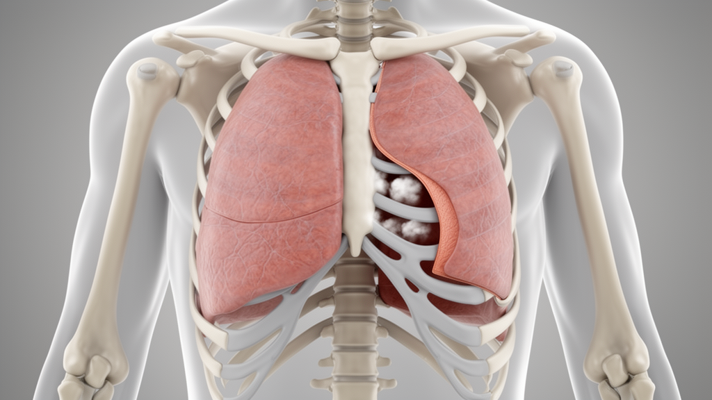
A comprehensive review published in Frontiers in Bioengineering and Biotechnology synthesizes current biomechanical understanding of pneumothorax, focusing on tissue failure mechanisms, computational simulation methods, and the path toward clinical translation. The study, led by researchers from The First Affiliated Hospital of Xi’an Jiaotong University, examines how damage to tissue structures responsible for maintaining pleural-cavity airtightness drives the development and persistence of the condition. By integrating continuum mechanics with clinical observations, the review outlines a framework for understanding the mechanical interactions among tissue failure, air leakage, pleural pressure changes, and global lung deformation.
The authors categorize pneumothorax-related tissue disruption into three idealized mechanical failure modes: stress-driven, material-driven, and overload-driven failure. Stress-driven failure occurs when thoracic geometry and local geometric discontinuities produce localized stress concentrations, particularly at the lung apex, which may explain the susceptibility to primary spontaneous pneumothorax in tall, slender individuals. Material-driven failure is associated with underlying pulmonary diseases such as emphysema or fibrosis, which alter tissue stiffness and load-bearing capacity, making the same external loading produce different stress responses compared to healthy tissue.
Overload failure results from external mechanical forces or abnormal pressure loading that exceeds local tissue capacity, a mechanism relevant to traumatic and iatrogenic pneumothorax. In traumatic cases, this involves penetrating or blunt forces, while in iatrogenic cases associated with mechanical ventilation, it involves excessive transpulmonary pressure and repetitive alveolar opening and closing. The review emphasizes that these modes are conceptual frameworks for interpreting tissue failure rather than established clinical classifications, as they often overlap and interact in actual disease processes.
Computational modeling is central to quantifying these mechanical processes, with the review detailing the application of hyperelastic, viscoelastic, and poroelastic constitutive models. Hyperelastic models, such as the Neo-Hookean, Mooney–Rivlin, and Ogden models, describe the nonlinear elastic behavior of lung parenchyma under finite deformation. These models rely on strain energy density functions to capture the complex stress-strain relationships of lung tissue, which cannot be adequately described by linear elastic models during large deformations like whole-lung collapse.
Viscoelastic models incorporate time-dependent behavior, accounting for hysteresis during cyclic loading and stress relaxation at constant strain. The review notes that lung parenchyma exhibits pronounced frequency-dependent behavior, with instantaneous mechanical responses varying with loading frequency. Generalized Maxwell models and fractional viscoelastic models are discussed as tools to capture these time-dependent characteristics, which are critical for simulating cyclic respiration and dynamic lung re-expansion.
For processes involving gas transport and solid deformation, poroelastic models couple the deformation of the solid lung skeleton with gas transport within the pore space. These models represent the lung as a two-phase porous medium, allowing for the simulation of bidirectional coupling where solid deformation alters local porosity and gas pressure, which in turn affects tissue deformation. The review highlights that while these models are powerful for studying ventilation and pressure-volume behavior, they require careful calibration of effective parameters such as porosity and permeability.
Fracture mechanics methods, including cohesive zone models (CZMs) and the extended finite element method (XFEM), are used to simulate tissue damage and crack propagation. CZMs define traction-separation laws along predefined interfaces, making them suitable for known fracture paths like pleural separation. XFEM uses enrichment functions to allow cracks to cross element boundaries, offering flexibility for uncertain crack paths. The review stresses that the choice of method depends on the specific failure mode, with continuum damage models often preferred for widespread tissue softening.
Despite these advanced modeling capabilities, the review identifies significant limitations in current research. A major constraint is the insufficient characterization of material properties, as lung tissue exhibits substantial interindividual variability and depends on disease state, age, and loading conditions. Homogeneous material parameters often fail to capture local mechanical variations, potentially affecting predictions of stress distributions. Additionally, the lack of rigorous experimental and clinical validation hinders the comparability of model predictions and their application to individualized clinical management.
The review discusses potential engineering applications, including patient-specific risk assessment based on biomechanical biomarkers. These biomarkers include anatomical features from imaging and mechanical indices derived from computational models, such as maximum principal stress and strain energy density. While these indices may help identify candidates for spontaneous pneumothorax, their clinical value requires prospective validation to demonstrate independent predictive value across different patient populations.
Intraoperative navigation and surgical planning are another area of potential application, where biomechanical models can characterize the non-rigid deformation of lung tissue from the inflated preoperative state to the collapsed intraoperative state. This could improve lesion localization during video-assisted thoracoscopic surgery (VATS) by reducing spatial misalignment between preoperative images and intraoperative anatomy. The review notes that while preliminary studies have shown promise in reducing localization errors, further evaluation of computational time and impact on surgical decision-making is needed.
The optimization of treatment strategies, particularly regarding the prevention of re-expansion pulmonary edema (RPE), is also explored. RPE is a rare but serious complication associated with rapid lung re-expansion, and its precise pathogenesis remains incompletely understood. Animal studies suggest that the duration of lung collapse and the rate of tissue deformation may contribute to injury, but quantitative evidence linking drainage conditions to mechanical lung injury is limited. Biomechanical models could provide a framework for comparing drainage conditions and identifying regions susceptible to excessive distension.
The review concludes that translating pneumothorax biomechanics into clinical practice requires establishing modeling frameworks with reliable material parameters and rigorous validation. Future research should prioritize the measurement of tissue properties, patient-specific model development, and multilevel validation against experimental and clinical data. Only when model reliability and clinical predictive value are progressively established can these computational methods provide a credible quantitative basis for preoperative risk assessment and treatment optimization.
The mechanical behavior of pneumothorax manifests across distinct physiological states defined by pressure dynamics and pathway patency. In closed pneumothorax, the air-leak pathway seals via local tissue apposition, blood clot formation, or elastic recoil, establishing a quasi-static equilibrium as trapped air resorbs. Open pneumothorax involves persistent communication between the pleural cavity and atmospheric air through a chest wall defect, eliminating transpulmonary pressure gradients. Tension pneumothorax acts as a dynamic emergency where a valve-like mechanism permits continuous air entry without egress, progressively escalating intrapleural pressure, compressing pulmonary vasculature, inducing mediastinal shift, and impairing cardiac output. Geometric quantification of the thoracic cage further clarifies structural vulnerability.
Characterizing the boundary behavior of the lung surface requires experimental evaluation of the visceral pleura. Planar biaxial tensile testing conducted by Ramirez and colleagues demonstrated that pleural membranes exhibit pronounced nonlinear elasticity, displaying progressive strain stiffening as collagen fibers are recruited under tension. While visceral pleura exhibits approximately isotropic in-plane mechanical properties across human, porcine, and rodent specimens, significant regional heterogeneity exists across lobes. Tissues excised from upper pulmonary lobes show distinct baseline and ultimate stiffness values compared with tissue from lower lobes. When regional material variations overlap with elevated apical stresses, localized microdamage, matrix remodeling, or tissue pore formation can occur. This interaction provides a biomechanical rationale for the predilection of primary spontaneous pneumothorax for apical subpleural regions, where mechanical load concentration coincides with regional tissue susceptibility.
Quantifying tissue failure requires distinguishing between ultimate material strength and empirical damage criteria. In direct evaluations of human lung tissue, Huang and colleagues conducted ex vivo uniaxial tensile tests on thirty-nine surgically resected bulla specimens and thirty-four parenchymal specimens. Across the samples failing within gauge length, the mean ultimate tensile strength reached 2.318 ± 1.541 megapascals for bulla tissue compared with 0.390 ± 0.210 megapascals for lung parenchyma. These measurements demonstrate marked disparities in load-bearing capacity, although the authors cautioned that uniaxial values cannot serve as universal in vivo failure criteria due to multiaxial physiological stress and non-uniform wall thickness. Conversely, strain-based damage thresholds derived from animal contusion models — such as principal strain limits of 3.5 percent, 8.8 percent, and 35 percent reported by Stitzel and colleagues, or 15.4 percent reported by Gayzik — represent computational associations with blunt trauma rather than universal crack-initiation criteria for spontaneous pneumothorax.
The mechanical consequences of therapeutic re-expansion similarly demand rigorous distinction between statistical clinical associations and underlying physical mechanisms. A meta-analysis of five retrospective cohort studies involving 1,093 patients by Willim and colleagues linked re-expansion pulmonary edema to longer symptom duration, with a pooled mean difference of 3.76 days, greater pneumothorax extent, and a history of smoking with an odds ratio of 1.94. Experimental animal models provide direct mechanistic insight into these clinical observations. Pavlin and Cheney demonstrated that applying negative suction of minus 100 Torr to rabbit lungs induced pulmonary edema in 85 percent of cases after seven to eight days of collapse, compared with 17 percent after three days and zero percent after thirty minutes. Furthermore, porcine mechanical ventilation experiments by Protti and colleagues documented that high tissue strain rates produced a 73 percent incidence of pulmonary edema compared to 20 percent under low strain rates. These findings indicate that pre-existing collapse duration, rapid suction pressure changes, and elevated strain rates collectively aggravate microvascular filtration and tissue injury during re-expansion.
Subject of Research: Biomechanical mechanisms and computational modeling of pneumothorax tissue failure and clinical translation
Article Title: Biomechanical analysis of pneumothorax: tissue failure, computational simulation, and clinical translation
Article References: Liu, A., Wang, T., Liang, R., Li, C., Xu, H., Ahmed, A.-A. W. H., Yu, C., Weng, Z., Fan, J., You, J., Cui, X., & Zhang, Y. (2026). Biomechanical analysis of pneumothorax: tissue failure, computational simulation, and clinical translation. Frontiers in Bioengineering and Biotechnology, 14, Article 1989437. https://doi.org/10.3389/fbioe.2026.1989437
Image Credits: AI Generated
DOI: 10.3389/fbioe.2026.1989437
Keywords: pneumothorax, biomechanics, tissue failure, computational modeling, finite element analysis, lung parenchyma, visceral pleura, constitutive models, re-expansion pulmonary edema, clinical translation, stress concentration, poroelasticity, fluid-structure interaction, surgical planning, pleural pressure
News Source: Gregory Coleman. (November 14, 2026). Review Maps Pneumothorax Biomechanics and Modeling Gaps. Scienmag.