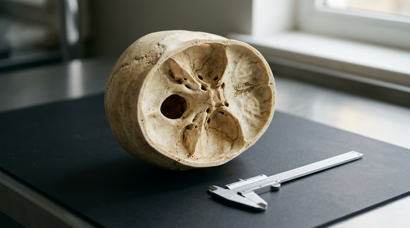
A forensic investigation evaluating the cranial anatomy of Egyptian adults has demonstrated that three-dimensional measurements of the sphenoid sinus provide measurable but modest predictive capacity when estimating biological sex and age. The cross-sectional study examined cone-beam computed tomography scans from two hundred and ninety adult individuals to evaluate whether internal skull cavities can serve as dependable anatomical markers when traditional skeletal remains are missing, damaged, or severely fragmented. Although the researchers identified statistically significant differences in dimensions between males and females as well as across defined age categories, statistical modeling demonstrated that the sphenoid sinus lacks the high discriminative power required for standalone forensic identification. Instead, the resulting predictive equations yielded an area under the receiver operating characteristic curve of 0.62 for biological sex classification and an age estimation error exceeding eleven years, establishing that these anatomical measurements are best suited as secondary, supplementary indicators within wider anthropological investigations.
Personal identification of human remains forms an essential component of medicolegal investigation, particularly in disaster victim identification, mass catastrophes, and forensic casework where recovered remains are fragmented, degraded, or incomplete. Establishing a biological profile routinely requires forensic anthropologists to determine biological sex, chronological age, stature, and population affinity from surviving skeletal elements. In contrast, the sphenoid sinus is situated deeply within the sphenoid bone at the central base of the cranium, structurally protected by surrounding cranial architecture from direct physical trauma and harsh environmental degradation. Because craniofacial morphology and pneumatization vary across human populations, investigators sought to establish dedicated baseline data for an Egyptian cohort, addressing a documented scarcity of population-specific morphometric standards.
The study protocol utilized archived radiological records collected from diagnostic imaging facilities in Cairo and Fayoum governorates, adhering to ethical standards approved by the Research Ethics Committee of the Faculty of Dentistry at Beni-Suef University. To ensure statistical power and balanced representation across the adult lifespan, the researchers applied convenience sampling to select scans from one hundred and forty-five male and one hundred and forty-five female patients aged eighteen to seventy-one years. The sample was systematically stratified into five distinct age cohorts comprising individuals aged eighteen to twenty-nine, thirty to thirty-nine, forty to forty-nine, fifty to fifty-nine, and sixty years or older, with exactly twenty-nine male and twenty-nine female scans assigned to each tier. Scans exhibiting developmental craniofacial anomalies, cranial fractures, evident pathological lesions, history of prior cranial surgery, or pronounced positioning misalignment were systematically excluded from the investigation.
The majority of imaging data was captured using a Planmeca ProMax 3D Mid system operating at standardized clinical exposure settings, including an operating voltage of ninety kilovolts peak, a current of eight milliamperes, a field of view measuring twenty by twenty centimeters, a voxel size of four hundred micrometers, a slice thickness of zero point five millimeters, and an exposure duration of thirteen point six seconds. Scans collected from Fayoum underwent sensitivity testing, which confirmed that their inclusion introduced no meaningful bias to the aggregate findings. Image data were exported in standard digital imaging communications format and evaluated within RadiANT viewing software under low-light conditions. Two experienced maxillofacial radiologists independently evaluated the scans using native orthogonal multiplanar reconstructions to preserve standardized orientation. Reliability testing confirmed excellent consistency, with intraclass correlation coefficients reaching 0.938 for intraobserver evaluation and 0.943 for interobserver agreement.
The radiological protocol quantified three standard anatomical distances across standardized planes: the latero-lateral distance measured as the widest span between the lateral walls on axial slices, the anteroposterior distance measured between the most anterior and posterior boundaries on sagittal views, and the inferior-superior distance captured between the lowest floor and highest roof on coronal reconstructions. Across the complete sample, the mean age was 43.15 years, with females averaging 42.40 years and males averaging 44.18 years. Descriptive statistics revealed that the latero-lateral diameter consistently represented the largest dimension, whereas the inferior-superior diameter exhibited the smallest values across both sexes. Male subjects demonstrated significantly greater mean values for the inferior-superior dimension at 25.84 millimeters compared to 24.88 millimeters in females, as well as greater latero-lateral dimensions at 37.43 millimeters compared to 35.24 millimeters. Conversely, females presented a slightly longer mean anteroposterior distance of 31.09 millimeters compared to 30.79 millimeters in males.
Analysis of dimensional patterns across progressive age groups revealed complex morphological dynamics throughout adult life. The inferior-superior measurement attained its highest average in individuals aged thirty to thirty-nine years at 27.21 millimeters, followed closely by the youngest cohort aged eighteen to twenty-nine years at 26.93 millimeters, before exhibiting a progressive reduction in older groups. The latero-lateral dimension peaked among individuals aged eighteen to twenty-nine years at an average of 40.37 millimeters and subsequently declined to its minimum of 33.35 millimeters in the forty to forty-nine age group, followed by minor expansions in subsequent decades. Conversely, the anteroposterior dimension showed an inverse trajectory, registering its lowest mean value of 28.82 millimeters among individuals aged forty to forty-nine years, but expanding substantially to 34.20 millimeters in the fifty to fifty-nine cohort and remaining elevated at 32.42 millimeters in subjects aged sixty and older, reflecting continuous late-stage anatomical variation.
Spearman rank correlation analyses demonstrated that dimensional variations tracked chronological age in statistically significant patterns across the population sample. Among male subjects, advancing age exhibited significant negative correlations with both the latero-lateral measurement at negative 0.532 and the inferior-superior distance at negative 0.486. In contrast, the anteroposterior measurement exhibited a significant positive correlation with age among females at 0.60 and across the entire pooled sample at 0.39. Regularization analyses using Lasso and Elastic Net regression retained all three linear parameters, confirming that each dimension provided non-redundant predictive information without harmful multicollinearity. Multivariable logistic regression revealed that the latero-lateral distance was the single statistically significant individual predictor for identifying male sex, generating an odds ratio of 1.08 with a ninety-five percent confidence interval spanning 1.02 to 1.14.
Despite statistical significance across specific anatomical dimensions, the resulting multivariable models demonstrated restricted practical accuracy. The cross-validated logistic regression model for biological sex classification yielded an area under the curve of 0.62, which corresponds to moderate discriminatory ability. Setting the optimal probability cutoff threshold at 0.49 via the Youden index produced a classification specificity of 0.76 and a sensitivity of 0.49, reflecting greater efficacy in correctly classifying male skulls than female crania, with an overall cross-validated accuracy of sixty-two percent. In forensic anthropology, conventional methodology generally demands classification accuracy approaching ninety-five percent under optimal conditions, while practical thresholds for compromised remains frequently expect posterior probabilities between seventy-five and eighty percent. Consequently, the observed sixty-two percent classification rate establishes that sphenoid sinus linear measurements cannot reliably establish sex in isolation.
Linear regression modeling developed to estimate chronological age similarly demonstrated clear limitations in individual predictive precision. Although all three linear dimensions emerged as statistically significant contributors to the regression equation, the combined multivariable formula accounted for only thirty-two percent of total variance in age, yielding a coefficient of determination of 0.32. The model exhibited a root mean squared error of 11.62 years, indicating that predicted ages deviate from actual chronological age by more than a decade on average. Forensic anthropologists observe that this degree of estimation uncertainty limits individual age reconstruction, as natural biological variability and age mimicry cause distinct individuals across varying chronological cohorts to exhibit overlapping sphenoid dimensions. The authors noted that while sinus pneumatization largely concludes during adolescence, ongoing bone remodeling and cranial base morphology continue to introduce subtle structural changes throughout adulthood.
The investigators emphasized several methodological constraints, noting that the cohort was recruited exclusively from Egyptian individuals, restricting the immediate application of derived equations to other ancestral groups without local validation. Additionally, the retrospective design meant scanner head alignment could not be actively standardized, and the software precluded full three-dimensional volumetric reorientation. The authors emphasized that three simple linear distances cannot fully capture the irregular internal architecture of the sphenoid cavity, suggesting that advanced volumetric segmentation and artificial intelligence tools may improve precision in future investigations. Nevertheless, because the deep cranial location of the sphenoid bone grants substantial protection against destruction, these documented morphometric benchmarks provide an objective supplementary reference for forensic specialists reconstructing biological profiles from heavily damaged or degraded cranial remains.
Subject of Research: Forensic evaluation of sphenoid sinus morphometry using cone-beam computed tomography for sex and age estimation in an Egyptian cohort
Article Title: Morphometric analysis of sphenoid sinus using cone-beam computed tomography: potential applications for age and sex estimation in an Egyptian population
Article References: Mohamad, N. E. E.-D., Hassan, F. M., Abo Elmagd, I. A., & Samir, W. (2026). Morphometric analysis of sphenoid sinus using cone-beam computed tomography: potential applications for age and sex estimation in an Egyptian population. Egyptian Journal of Forensic Sciences, 16(1), Article 45. https://doi.org/10.1186/s41935-026-00540-x
Image Credits: AI Generated
DOI: 10.1186/s41935-026-00540-x
Keywords: sphenoid sinus, cone-beam computed tomography, forensic anthropology, biological profiling, sex estimation, age estimation, cranial morphometry, logistic regression, multiple linear regression, Egyptian population, radiology, paranasal sinuses, receiver operating characteristic, skeletal identification, sexual dimorphism, interobserver reliability
News Source: Peter Mason. (November 13, 2026). Sphenoid Sinus Dimensions Offer Modest Forensic Value for Estimating Sex and Age. Scienmag.