Researchers have shown that deep learning models applied to standard light microscopy can identify distinct metabolic states of the human gut bacterium Clostridium scindens linked to the production of deoxycholic acid, a genotoxic secondary bile acid implicated in colorectal cancer. Colorectal cancer is among the most prevalent malignancies globally, and early-stage screening remains critical because symptoms often do not manifest during the initial phases of oncogenesis. Emerging scientific evidence implicates the gut microbiome as an active driver of colorectal cancer development, inspiring efforts to engineer microbiome-based diagnostic tools. Gut microorganisms influence individual oncological risk through metabolic pathways, generating either protective compounds or carcinogenic agents within the intestinal tract. Clostridium scindens converts cholic acid into deoxycholic acid, a pro-inflammatory metabolite that promotes malignant transformation. While mass spectrometry and genomic sequencing can quantify these metabolites, their substantial cost and operational complexity hinder wide clinical deployment, underscoring the appeal of accessible light microscopy.
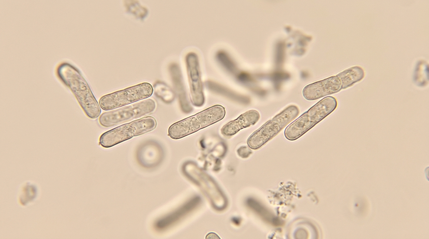
To assess whether computational computer vision frameworks can detect bacterial metabolic states directly from visual features, the investigators cultured Clostridium scindens anaerobically at 37 degrees Celsius for 24 hours across four defined experimental conditions. Clostridium scindens cultured in rich media alone represented the non-producing, health-associated baseline state, whereas cultures supplemented with 100 micromolar cholic acid provided the necessary biochemical precursor to induce deoxycholic acid production, representing the cancer-associated state. Two additional experimental conditions evaluated Clostridium scindens in anaerobic co-culture with either Bacteroides thetaiotaomicron or Bacteroides vulgatus in the presence of 100 micromolar taurocholic acid, an upstream conjugated bile acid that Bacteroides species convert into cholic acid. Overnight bacterial cultures with an optical density of approximately 0.2 at 600 nanometers were harvested, deposited onto glass slides using a CytoSpin centrifuge at 500 revolutions per minute for five minutes, flame-fixed, Gram-stained, mounted, and imaged at 100x magnification using the TissueFAX automated microscopy system with 0.2-micrometer z-stack depth.
The research team developed and evaluated three distinct computational modeling methodologies to process the resulting bacterial imaging data: whole-image classification, automated single-cell detection followed by classification, and semantic pixel-level image segmentation. Across all analytical pathways, microscopic image files were imported into memory and standardized using the ImageDataGenerator class from TensorFlow’s Keras application programming interface, which normalized pixel values and partitioned the data into an 80 percent training set and a 20 percent testing set. Within the training allocation, 25 percent of the image files were reserved for internal validation routines during model optimization. All images were formatted to dimensions of 224 by 224 pixels to match the input tensor specifications of the convolutional neural network pipelines. Ground-truth experimental class labels were assigned automatically based on directory structures designating the specific presence or absence of cholic acid supplementation as well as the bacterial species present in each culture condition.
For whole-image classification, the authors evaluated three neural network strategies to establish how model complexity and pre-training affected diagnostic accuracy. They initially constructed a custom convolutional neural network from scratch using TensorFlow and Keras, employing three convolutional layers with 32, 64, and 128 filters of 3 by 3 kernel dimensions and Rectified Linear Unit activations, coupled with 2 by 2 max-pooling layers, a flattening layer, an intermediate dense layer of 128 neurons, and a single sigmoid output neuron. Next, the team deployed transfer learning by taking pre-trained ResNet50 and DenseNet architectures trained on ImageNet, freezing their convolutional bases to serve as fixed feature extractors while appending identical custom classification heads. Finally, they developed an enhanced ResNet50 model by fine-tuning the ImageNet-initialized network on the Digital Images of Bacterial Species repository, an external microbiological dataset comprising 33 distinct bacterial species, allowing the convolutional base to learn specialized representations of diverse bacterial morphologies prior to target training.
Empirical whole-image evaluations revealed that pre-trained networks substantially surpassed the custom convolutional neural network, with the microbial-enhanced model delivering the highest overall classification accuracy. When distinguishing Clostridium scindens monocultures cultured with cholic acid from those cultured in media alone, the custom de novo network achieved an accuracy of only 0.34 and an F1 score of 0.39. In comparison, DenseNet demonstrated an accuracy of 0.75 with an F1 score of 0.85, whereas standard ResNet attained 0.85 accuracy and an F1 score of 0.91. Integrating the Digital Images of Bacterial Species dataset yielded an accuracy of 0.89 with a standard deviation of 0.0056, an F1 score of 0.90, and a receiver operating characteristic area under the curve of 0.85. Mann-Whitney U testing confirmed that this bacterial-enhanced ResNet demonstrated statistically superior accuracy over standard ResNet (p = 0.037), DenseNet (p = 0.0063), and the custom network (p = 0.00021), while also leading co-culture classification with 0.86 accuracy.
To overcome the limitations of whole-field evaluations, the investigators implemented a two-step framework for individual cell detection and automated classification. Cell detection was conducted using Otsu and adaptive thresholding methods to isolate individual bacterial cells across variable grayscale image fields without requiring manual human annotation. The team tested multiple values for the adaptive subtraction constant C, ranging from 1 to 8, on images of Clostridium scindens cultured without cholic acid to optimize segmentation parameters. Setting C = 1 generated excessive sensitivity, detecting 8,375 putative cells alongside non-cellular noise, yielding an F1 score of 0.81. Setting C = 3 achieved the optimal balance between sensitivity and specificity, isolating 9,252 cells with 0.91 precision, 0.86 recall, and a peak F1 score of 0.89. Higher constant values progressively omitted valid targets, with C = 4 isolating 7,180 cells (F1 score 0.70), C = 5 detecting 5,500 cells (F1 score 0.52), and C = 8 identifying 2,050 cells (F1 score 0.25).
Following automated boundary detection at the optimal C = 3 configuration, the extracted single-cell image patches were classified using a ResNet50 model trained for 30 epochs with batch sizes of 16 and random rotation augmentations. In monoculture experiments without cholic acid, the training set contained 35,990 total cells from 20 images, yielding 27,353 detected Clostridium scindens cells, of which 23,685 were correctly classified. During internal validation across five images containing 13,806 cells, 8,622 cells were detected, with 6,439 cells (74 percent) correctly identified as non-producing cells and 2,183 (26 percent) misclassified. On an independent external test set of five images containing 13,559 total cells, 8,405 cells were extracted, with 5,622 cells (66 percent) correctly classified and 2,783 (34 percent) misclassified. For cholic acid-supplemented monocultures, the model was trained on 5,398 detected cells from 20 images, achieving an internal validation accuracy of 69 percent and an independent external testing accuracy of 62 percent.
To identify the specific visual features guiding neural network decisions, the research team implemented Gradient-weighted Class Activation Mapping across the trained classification models. This interpretability technique computes the gradient of target class output scores with respect to convolutional feature map activations, producing heatmaps where warmer red and yellow color spectra highlight the most influential spatial regions within each microscopic field. While deoxycholic acid synthesis occurs biochemically within intracellular enzymatic pathways, the class activation maps revealed that the models did not prioritize internal cellular regions. Instead, the heatmaps consistently localized critical discriminative features directly along the bacterial cell membrane and within the immediate surrounding optical halo region.
The researchers next tested the trained per-cell classifier in an unsupervised setting using mixed co-cultures containing Clostridium scindens alongside non-carcinogenic Bacteroides species. Liquid chromatography-mass spectrometry verified that co-cultures containing Bacteroides vulgatus produced substantially higher deoxycholic acid concentrations (6.89 × 10^6 ± 2.55 × 10^5 nanograms per milliliter) than co-cultures containing Bacteroides thetaiotaomicron (1.61 × 10^5 ± 5.49 × 10^4 nanograms per milliliter). When evaluating 1,538 cells from 20 test images of the low-producing Bacteroides thetaiotaomicron condition, the system detected 529 of 1,021 Clostridium scindens cells (a 51.8 percent detection rate) and classified 72.2 percent (382 cells) as non-producing. Conversely, across 1,958 cells from 20 images of the high-producing Bacteroides vulgatus condition, the model detected 705 of 1,372 Clostridium scindens cells (a 51.4 percent detection rate) and categorized 56.2 percent (396 cells) as carcinogen-producing, demonstrating that single-cell predictions reliably reflected verified differences in biochemical carcinogen output.
To benchmark semantic segmentation, the investigators trained nnU-Net, a specialized medical imaging framework featuring symmetrical five-block two-dimensional convolutional pathways with instance normalization, skip connections, and a combined cross-entropy and Dice loss function. Across 50 training images with fivefold cross-validation and 25 external test images, nnU-Net achieved Dice similarity coefficients of 98 percent in validation and 87 percent in testing for Clostridium scindens, alongside testing Dice scores of 76 percent for Bacteroides vulgatus and 74 percent for Bacteroides thetaiotaomicron, with detection accuracies spanning 0.87 to 0.90 for Clostridium scindens and 0.80 for both Bacteroides species. The authors noted that detection rates in mixed co-cultures were constrained by conventional adaptive thresholding, suggesting that future improvements should incorporate higher-resolution microscopy, unified end-to-end architectures such as You Only Look Once, and transformer-based attention modules. Dataset images are available in the EMBL-EBI BioImage Archive (S-BIAD1671) and code is hosted on GitHub.
Subject of Research: Deep learning imaging analysis of bacterial metabolic states linked to carcinogen production
Article Title: Deep learning imaging analysis to identify bacterial metabolic states associated with carcinogen production
Article References: Orouskhani, M., Rauniyar, S., Morella, N., Lachance, D., Minot, S. S., & Dey, N. (2025). Deep learning imaging analysis to identify bacterial metabolic states associated with carcinogen production. Discover Imaging, 2(1), Article 2. https://doi.org/10.1007/s44352-025-00006-1
Image Credits: AI Generated
DOI: 10.1007/s44352-025-00006-1
Keywords: Deep Learning, Gut Microbiome, Clostridium scindens, Colorectal Cancer, Deoxycholic Acid, Microscopy, Computer Vision, nnU-Net, Deep, learning, imaging, analysis
News Source: Blake Davidson. (November 13, 2026). Deep Learning Identifies Carcinogen-Producing Gut Bacteria From Light Microscopy Images. Scienmag.