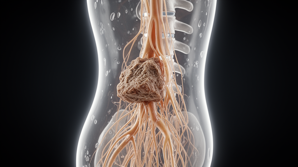
The adverse event occurred following an endoscopic decompression and discectomy procedure performed for an initially unrecognized intradural lumbar disc herniation. The case demonstrates that unretrieved intradural disc material can mobilize within the spinal canal after endoscopic intervention, leading to acute secondary neurological deterioration.
The patient presented to medical care with a clinical history of recurrent low back pain accompanied by progressively worsening numbness and radicular pain in her right leg. Preoperative diagnostic imaging demonstrated a giant central disc herniation located at the L3–4 level that caused marked compression of the cauda equina.
To address the significant neural compression, surgeons performed an endoscopic decompression and discectomy utilizing a right-sided L3/4 interlaminar surgical approach. During this endoscopic intervention, the surgical team identified a longitudinal ventral dural tear along with a free intradural nucleus pulposus fragment. The operating team removed the visible intradural fragment through the endoscopic working channel before completing the procedure.
In the immediate postoperative period, the patient reported relief and improvement in her right-sided radicular pain. However, a distinct neurological deficit soon emerged, with distal motor dysfunction of the left lower extremity presenting as the dominant postoperative complication. This motor impairment was rapidly followed by the onset of perineal hypoesthesia and saddle hypoesthesia, signaling progressive cauda equina compromise. Prompted by these acute neurological findings, clinicians conducted an emergency magnetic resonance imaging scan, which revealed a distinct compressive lesion located posterior to the L4 vertebra.
Faced with evidence of persistent canal compromise, the clinical team executed a second-stage open microscopic exploration. During this second procedure, surgeons confirmed and removed a residual intradural nucleus pulposus fragment that had undergone delayed caudal displacement below the initial operative site. Following this microscopic removal, the patient demonstrated neurological and functional improvement; nevertheless, both motor function and bowel recovery remained incomplete when evaluated at the six-month postoperative milestone.
The report underscores that intradural lumbar disc herniation represents a rare pathological entity that is notoriously difficult to diagnose in preoperative assessments and easily mistaken for giant central disc herniations. The study authors recommend that surgeons actively consider the possibility of intradural disc herniation whenever evaluating giant central disc herniations that present with marked cauda equina compression, particularly when conventional magnetic resonance imaging results are inconclusive. In such complex diagnostic presentations, the authors suggest that high-resolution or contrast-enhanced magnetic resonance imaging may provide critical diagnostic clarification.
Regarding operative management, the authors note that endoscopic treatment of suspected intradural disc herniation can be feasible under circumstances where the dural opening is strictly controlled and surgical visualization remains fully adequate. Nonetheless, they highlight that continuous fluid irrigation during endoscopy represents one of several possible contributing mechanisms leading to the unintended hydrodynamic displacement and caudal migration of residual intradural disc fragments. To mitigate this surgical risk, the authors emphasize that if complete intradural exploration or the reliable exclusion of residual disc fragments cannot be successfully achieved through the endoscopic corridor, surgeons should consider timely conversion to open microscopic exploration.
Subject of Research: Delayed caudal migration of residual intradural disc fragment causing acute cauda equina dysfunction
Article Title: Delayed caudal migration of a residual intradural disc fragment after endoscopic discectomy for unrecognized intradural disc herniation causing acute cauda equina dysfunction: a case report
Article References: Ren, Z., Lv, L., Wang, J., Lu, J., & Zhang, X. (2026). Delayed caudal migration of a residual intradural disc fragment after endoscopic discectomy for unrecognized intradural disc herniation causing acute cauda equina dysfunction: a case report. BMC Surgery. https://doi.org/10.1186/s12893-026-04257-5
Image Credits: AI Generated
DOI: 10.1186/s12893-026-04257-5
Keywords: intradural lumbar disc herniation, cauda equina dysfunction, delayed caudal migration, endoscopic discectomy, residual disc fragment, nucleus pulposus, ventral dural tear, interlaminar approach, microscopic exploration, continuous irrigation, emergency MRI, contrast-enhanced MRI, high-resolution MRI, saddle hypoesthesia, perineal hypoesthesia, motor dysfunction, bowel recovery
News Source: Ophelia Keating. (November 12, 2026). Residual Intradural Disc Fragment Migration Triggers Acute Cauda Equina Dysfunction. Scienmag.