A comparative microscopic evaluation of human hair fibers has revealed that excessive cosmetic bleaching induces severe structural breakdown across both the outer protective cuticle and the interior cortex. Using scanning electron microscopy and high-magnification transmission electron microscopy, researchers observed that repeated exposure to alkaline bleaching mixtures stripped away overlapping surface scales and left behind adhered endocuticle debris, while entirely removing melanin granules within the cortex. In samples subjected to three consecutive bleaching cycles, the outer cuticle layer experienced extensive mechanical delamination, exposing the underlying cortical architecture to direct environmental contact. While the natural microfibrillar architecture within individual macrofibrils retained its recognizable fingerprint-like alignment, the loss of cortical mass and cellular cohesion demonstrated how aggressive chemical lightening fundamentally compromises the mechanical and physical integrity of human hair.
Cosmetic hair bleaching relies on an irreversible chemical reaction designed to alter natural pigmentation by destroying chromophores embedded within cortical melanin granules. The experimental protocol utilized healthy, untreated brown hair collected from a female donor in her twenties who had no prior history of chemical treatments, such as dyeing, bleaching, or perming. To simulate intensive salon lightening, the researchers applied a standard formulation composed of an alkaline bleaching powder with a pH between 9 and 11 combined in a one-to-one ratio with a six percent hydrogen peroxide oxidizing agent from Blondor, Wella Korea. The prepared mixture was applied approximately one centimeter away from the hair root. To accelerate the oxidative chemical reaction, thermal energy was applied using a hair dryer positioned approximately twenty centimeters from the strands for one to two minutes. The treated fibers were then maintained at room temperature for twenty minutes before being thoroughly rinsed with running tap water and allowed to air dry, a cycle repeated three times.
Following three bleaching cycles, optical microscopy confirmed a complete visual transition from the donor’s natural brown baseline to a bright yellow hue, reflecting the extensive degradation of natural pigment. To prepare these specimens for scanning electron microscopy, both normal and bleached strands were sectioned into one-millimeter fragments with a double-edged razor. The specimens underwent pre-fixation in two-and-a-half percent paraformaldehyde-glutaraldehyde buffered at pH 7.4 in phosphate buffer at four degrees Celsius, followed by two separate rinses in the same buffer solution. Post-fixation was carried out using one percent osmium tetroxide in phosphate buffer at four degrees Celsius for one hour. After secondary washing, samples were systematically dehydrated through an ascending ethanol series, transferred into isoamyl acetate, and processed through a critical point dryer. Dried hair shafts were subsequently coated with a twenty-nanometer platinum layer using an ion coater before being examined under a scanning electron microscope operated at an accelerating voltage of fifteen kilovolts.
For ultra-high-resolution transmission electron microscopy of internal cell components, separate hair segments measuring three millimeters in length were processed through an identical double-fixation routine in glass bottles, using two fifteen-minute washes in 0.4 molar phosphate buffer following primary aldehyde fixation. After post-fixation in one percent osmium tetroxide and complete ethanol dehydration, the specimens were transitioned through propylene oxide and embedded in an Epon-Araldite resin mixture. Polymerization was conducted in a specialized vacuum drying oven set to sixty degrees Celsius over a continuous thirty-six-hour period. Embedded blocks were initially sectioned into one-micrometer semi-thin slices using an ultramicrotome and stained with toluidine blue for preliminary light microscopy. For nanoscale structural examination, ultra-thin sections were sliced, collected on copper grids, double-stained with uranyl acetate and lead citrate, and imaged on a transmission electron microscope operating at one hundred kilovolts.
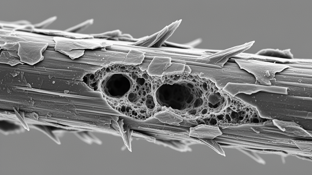
Scanning electron micrographs of untreated normal hair revealed a predominantly intact, smooth outer envelope characterized by tightly layered cuticle scales with gently rounded exposed margins. Although high-magnification scanning of normal fibers identified minor surface imperfections, such as slight edge irregularity and localized scale separation attributed to everyday friction and routine washing, the overall protective sheath remained contiguous and continuous. In contrast, bleached hair exhibited extensive surface weathering marked by brittle, fractured, and detached cuticle scales. At higher magnifications, the outermost cuticle scales were seen to peel completely away from underlying cell layers. Fragmented remnants of endocuticle debris remained adhered to the newly exposed cuticle surfaces, producing a jagged, irregular topography. In regions exhibiting the most pronounced damage, the entire cuticle sheath was sheared away, laying bare the cortical surface and generating prominent longitudinal fissures running parallel to the fiber axis.
Cross-sectional transmission electron microscopy of normal hair clearly demonstrated the multi-layered organization of intact cuticle cells, which were structurally differentiated into an outer exocuticle and an inner endocuticle based on electron density. In these untreated specimens, the endocuticle displayed higher electron density than the adjacent exocuticle and contained occasional natural micro-holes measuring between 0.1 and 0.5 micrometers in diameter, while the exocuticle remained dense, smooth, and free of physical defects. Within the normal cortex, cortical cells were densely packed in irregular, polygonal profiles. These cells contained tightly bundled, elliptical macrofibrils measuring roughly 0.5 to 1.0 micrometer across, surrounded by cell membrane complexes approximately twenty nanometers thick. Crystalline microfibrils within these macrofibrils formed regular, fingerprint-like concentric arrays embedded in an amorphous sulfur-rich protein matrix, while intact, oval melanin granules measuring approximately 0.5 micrometers in transverse diameter lined the inter-macrofibrillar spaces.
Transmission electron microscopy of the bleached fibers uncovered widespread internal disruption, most notably the near-total detachment of remaining cuticle cells from the underlying cortical boundary. The most dramatic microstructural alteration occurred within the cortex itself, where intact, electron-dense melanin granules were completely absent. Instead, the spaces formerly occupied by melanin granules were converted into large, hollow pores and irregularly shaped cavities. The investigators noted that alkaline oxidation had solubilized the granule matrices, leaving only sparse, undissolved granular remnants clinging to the perimeter walls of these newly created internal voids. Although the macrofibrils themselves preserved their overall elliptical shape and internal fingerprint-like microfibrillar organization, the surrounding cytoplasm exhibited pronounced structural deformation. Lysed cellular remnants originating from destroyed nuclei and cellular organelles were scattered throughout the cortex, distinguished by their irregular shapes and high electron density.
The study highlights how the chemical mechanics of persulfate-based bleaching agents disrupt hair integrity through multiple interactive pathways. Concurrently, the formulation extracts endogenous lipids, particularly 18-methyleicosanoic acid situated within the epicuticle layer. The depletion of this surface lipid barrier dramatically increases fiber permeability, facilitating rapid inward diffusion of high concentrations of hydrogen peroxide into the cortex. Within the cortical cells, the chemical reagents target the melanin granules, breaking down chromophoric chemical bonds through an alkaline oxidation process that dissolves the granule core. This destructive chemical cascade not only eliminates pigmentation but also leaves the interior cortex riddled with empty cavities, weakening the cohesive matrix that binds the structural macrofibrils together.
The authors contextualized their microscopic observations against earlier hair research, noting important points of concordance as well as notable experimental divergences. While previous literature reported by Kuzuhara had indicated that bleaching black human hair caused a detectable decrease in endocuticle electron density alongside an increase in exocuticle electron density, the present study observed no measurable shifts in electron density between these cuticle sublayers. Instead, the current investigation revealed a marked increase in the total frequency and size of holes within the endocuticle of bleached hair relative to normal controls.
The investigators concluded that excessive cosmetic bleaching severely undermines the physical stability of human hair by dismantling its protective exterior and perforating its internal architecture. The complete loss of protective cuticle scales and the formation of longitudinal fissures allow external moisture from washing and humidity to penetrate directly into the cortex. Because macrofibrils within the cortex degrade further upon sustained contact with environmental moisture, cortical porosity directly accelerates fiber weathering. Furthermore, because tensile strength and elasticity are primarily generated by alpha-keratin complexes within the cortex, the void spaces created by dissolved melanin granules and damaged disulfide bonds render bleached fibers highly vulnerable to split ends and spontaneous breakage. These ultrastructural findings demonstrate that repeated chemical lightening causes irreversible damage to both the surface cuticle and the deep cortex, emphasizing the structural trade-offs inherent in aggressive cosmetic hair treatments.
Subject of Research: Electron microscopic analysis of hair cuticle degradation and cortical microstructural damage caused by excessive bleaching
Article Title: Effects of excessive bleaching on hair: comparative analysis of external morphology and internal microstructure
Article References: Kim, D. H., Oh, S. H., & Chang, B. S. (2024). Effects of excessive bleaching on hair: comparative analysis of external morphology and internal microstructure. Applied Microscopy, 54(1), Article 11. https://doi.org/10.1186/s42649-024-00104-0
Image Credits: AI Generated
DOI: 10.1186/s42649-024-00104-0
Keywords: Hair bleaching, Transmission electron microscopy, Scanning electron microscopy, Hair cuticle, Hair cortex, Melanin granules, Macrofibrils, Microfibrils, Keratin degradation, Cuticle scale detachment, Cortical porosity, Hydrogen peroxide, Endocuticle, Exocuticle, Hair weathering, Longitudinal fissures, Disulfide bonds
News Source: Denise Maddox. (November 11, 2026). Electron Microscopy Shows Excessive Bleaching Strips Cuticle Layers and Cavitates Hair Cortex. Scienmag.