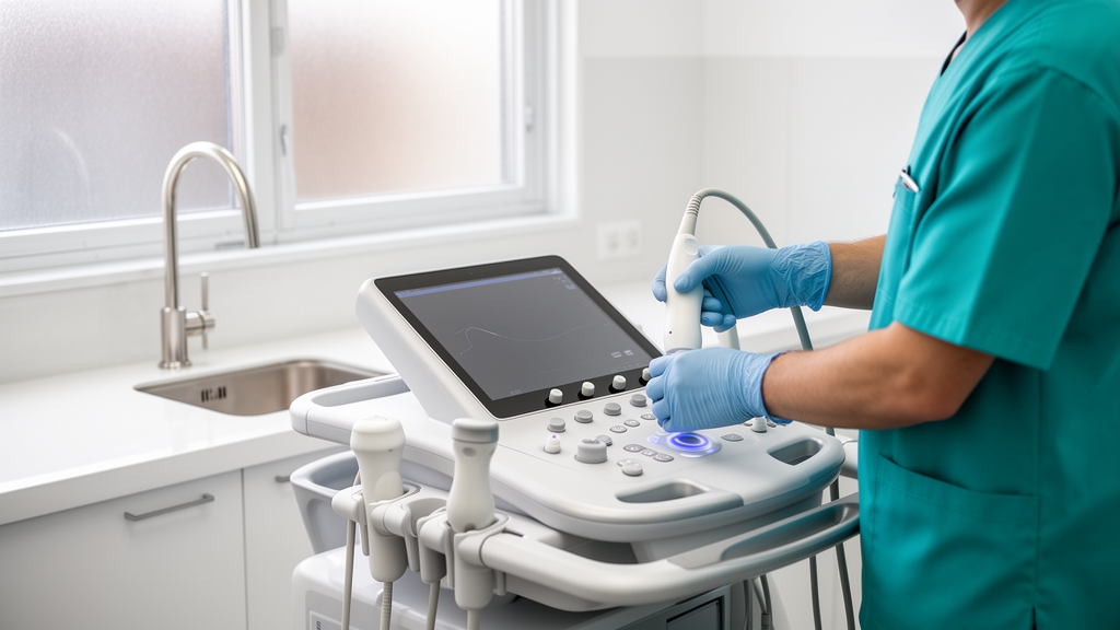
A systematic review and meta-analysis published in Basic and Clinical Andrology demonstrates that varicocele substantially alters both the physical stiffness and anatomical volume of testicular tissue. Varicocele, recognized as an abnormal dilation and tortuosity of the pampiniform plexus veins within the spermatic cord, represents a primary vascular disorder and a major contributor to both primary and secondary male infertility. While conventional color duplex ultrasound remains the standard clinical tool for confirming the presence of venous reflux and dilation, it lacks the technical capacity to detect parenchymal degradation prior to the development of overt testicular atrophy. To evaluate subtle biomechanical changes in testicular parenchyma, researchers led by Lihong Wang, Xiaoqi Huang, and Yinman Ding synthesized data from published clinical trials employing shear wave elastography. This advanced non-invasive imaging modality estimates tissue elasticity by measuring the propagation velocity of acoustic shear waves passing through the parenchyma. The quantitative synthesis confirms that varicocele-affected testes exhibit significantly elevated stiffness compared to both unaffected contralateral testes and healthy control organs, while also displaying a distinct reduction in organ volume.
The clinical scope of varicocele is extensive, affecting approximately 15 percent of adult males in the general population. Among men presenting with primary infertility, the condition is diagnosed in 35 to 44 percent of cases, and this prevalence escalates to between 45 and 81 percent among men experiencing secondary infertility. Although some individuals with varicocele retain natural fertility and conceive without medical intervention, the condition frequently causes progressive, time-dependent deterioration of testicular function, hypofunction, localized discomfort, and scrotal pain. The underlying physiological mechanisms driving testicular injury involve complex vascular and cellular disturbances. In affected individuals, the pampiniform plexus veins undergo pronounced dilatation, stasis, and heightened intravascular pressure. Associated pathological processes include elevated scrotal temperature, hypoxia, reduced testicular blood flow, androgen deprivation, and the retrograde reflux of toxic renal and adrenal metabolites into the testicular microcirculation. These cumulative insults promote oxidative stress, DNA damage, and apoptosis within germ cells, leading to microvascular intimal hyperplasia, endothelial cell degeneration, spermatogenic cell shedding, peritubular fibrosis, and interstitial edema.
To provide clear quantitative evidence regarding these parenchymal alterations, the investigators executed a systematic literature search in June and July 2024 across major biomedical databases, including PubMed, EMBASE, and MEDLINE, without imposing language or geographic limitations. Eligible clinical trials were required to incorporate comparative designs contrasting patients diagnosed with varicocele against control cohorts, provide verifiable numerical data on testicular stiffness and volume reported as means and standard deviations or medians and quartiles, and provide full-text accessibility. Studies that failed to distinguish between ipsilateral varicocele-affected testes and contralateral testes were systematically excluded from consideration. After screening an initial retrieval pool of 87 articles, the authors eliminated 41 publications following preliminary abstract and title screening, 21 studies for lacking proper anatomical distinction between ipsilateral and contralateral sides, and four studies due to insufficient data. Ultimately, seven observational clinical trials encompassing 1,118 individual testes met all rigorous inclusion criteria, representing clinical research conducted across Spain, Turkey, Italy, Poland, and China.
Quality assessment for the included investigations was conducted independently by two reviewers using the Preferred Reporting Items for Systematic Reviews and Meta-analyses framework. Because inherent biological characteristics and exposure to risk factors cannot be ethically or practically randomized in human subjects, the meta-analysis drew evidence from comparative clinical trials and observational cohorts. Studies were categorized as having a low risk of bias when all methodological quality requirements were fulfilled, or moderate risk when one or more quality standards remained unclear or partially satisfied. The extracted data included publication year, first author, participant age, country of origin, and absolute sample sizes, which ranged from 13 to 116 testes per study. Statistical synthesis was performed using Review Manager version 5.4, calculating mean differences with corresponding 95 percent confidence intervals for continuous outcomes. Heterogeneity across individual trial outcomes was quantified using the I-squared statistic, with random-effects models applied whenever heterogeneity exceeded 50 percent and fixed-effects models utilized when heterogeneity remained at or below that threshold.
The primary outcome analysis evaluated changes in tissue stiffness measured via shear wave elastography in kilopascals. When comparing varicocele-affected ipsilateral testes to contralateral testes across all seven included studies representing 718 organs, the meta-analysis revealed significant heterogeneity, necessitating a random-effects model. The pooled mean difference showed an increase in stiffness of 0.93 kilopascals on the affected side, with a 95 percent confidence interval spanning from 0.34 to 1.52 kilopascals and a statistically significant difference marked by p less than 0.05. A parallel analysis across five studies examining 660 testes compared the varicocele-affected group directly with healthy control testes from unaffected subjects. Applying a random-effects model due to inter-study variability, the pooled mean difference was 1.07 kilopascals, with a 95 percent confidence interval of 0.03 to 2.12 kilopascals, again confirming a statistically significant elevation in stiffness in affected testes compared to healthy controls at p less than 0.05.
A particularly intriguing finding emerged from the biomechanical assessment of the contralateral, non-varicocele-bearing testes in comparison to normal control testes. Drawing on data from five studies comprising 634 testes, the evaluation demonstrated an absence of heterogeneity, allowing the application of a fixed-effects statistical model. The resulting pooled mean difference was minus 0.12 kilopascals, with a tight 95 percent confidence interval between minus 0.23 and minus 0.02 kilopascals, demonstrating that the stiffness of contralateral testes was significantly lower than that of normal healthy controls at p less than 0.05. This observation highlights that the systemic or hemodynamic impact of unilateral varicocele does not leave the opposite gonad entirely unaffected. However, rather than developing the heightened rigidity characteristic of the ipsilateral side, the contralateral testis displays a slight reduction in tissue elasticity relative to healthy baseline controls, illustrating complex bilateral physiological alterations that warrant further investigation.
Regarding secondary outcomes, the meta-analysis systematically evaluated changes in testicular volume measured in milliliters. Across seven studies analyzing 718 testes, the comparison between varicocele-affected ipsilateral testes and contralateral testes demonstrated substantial heterogeneity. Under a random-effects model, the pooled mean difference was minus 2.30 milliliters, with a 95 percent confidence interval ranging from minus 3.96 to minus 0.64 milliliters and a significant difference at p less than 0.05, establishing that affected testes were measurably smaller than contralateral organs. Similarly, when five studies comprising 660 testes compared affected ipsilateral testes to normal control organs, a random-effects synthesis yielded a pooled mean difference of minus 2.50 milliliters, with a 95 percent confidence interval from minus 4.04 to minus 0.96 milliliters at p less than 0.05. In stark contrast, when five studies analyzing 634 testes compared contralateral testes directly with normal controls, a fixed-effects model identified a pooled mean difference of minus 0.48 milliliters, with a confidence interval spanning minus 1.09 to 0.13 milliliters, indicating no statistically significant difference in volume at p greater than 0.05.
The discordance between objective elastography measurements and traditional physical examination reveals a critical clinical paradox. Patients presenting with varicocele frequently describe the affected testicle as feeling noticeably softer during manual scrotal palpation. However, quantitative shear wave elastography establishes that the actual internal parenchyma of the diseased testis is significantly more rigid than healthy tissue. This tactile discrepancy arises because degenerative changes in germ cells, parenchymal atrophy, and apoptotic cellular loss can cause a loss of external turgor, whereas microscopic microvascular intimal hyperplasia, endothelial degeneration, and progressive interstitial fibrosis progressively stiffen the internal tissue architecture. Histopathological investigations in both human specimens and animal rabbit models corroborate that varicocele induces peritubular fibrosis, Leydig cell atrophy, and a reduction in seminiferous tubule diameter. Because acoustic shear waves travel at accelerated speeds through denser, fibrotic structures and slower speeds through soft tissues, elastography reliably maps this microscopic stiffening that manual palpation fails to detect.
Importantly, the meta-analysis demonstrated that the observed volumetric reduction in varicocele-affected testes does not correlate directly with quantitative shear wave elasticity values. This absence of linear correlation indicates that macroscopic organ volume cannot serve as an accurate or reliable surrogate for assessing the underlying severity of parenchymal injury or interstitial fibrosis. Although testicular biopsy represents the definitive histological standard for characterizing parenchymal damage and seminiferous tubule pathology, its invasive nature precludes routine clinical deployment in infertile men. Routine semen analysis provides indirect functional information but cannot visualize structural architecture. Consequently, quantitative elastography values, particularly when expressed in kilopascals rather than meters per second, offer superior diagnostic power, as prior research demonstrates a larger area under the receiver operating characteristic curve for kilopascal metrics. Elevated stiffness values directly reflect advancing interstitial fibrosis, positioning elastography as an objective diagnostic tool capable of monitoring subclinical progression or post-surgical recovery.
Despite these compelling conclusions, the authors outline several methodological limitations that temper the clinical interpretation of the pooled data. The overall synthesis was constrained to seven published investigations incorporating a total of 1,118 testes, with individual study cohorts remaining relatively modest in size. The reliance on published observational comparative trials and non-randomized clinical cohorts introduces potential selection biases and limits statistical power to detect subtler subgroup interactions. Additionally, variations in ultrasound equipment, differing technical protocols, distinct operator skill levels across trial centers, and the omission of unpublished gray literature could have introduced heterogeneity into the pooled estimates. To resolve these challenges and validate shear wave elastography as a routine staging and monitoring standard in andrological practice, the researchers emphasize that large-scale, multicenter prospective randomized controlled trials will be essential to establish definitive elastographic thresholds for clinical decision-making.
Subject of Research: Shear wave elastography assessment of testicular stiffness and volume in varicocele
Article Title: Shear wave elastography in assessing stiffness and volume of varicocele-affected and normal testes: a systematic review and meta-analysis
Article References: Wang, L., Huang, X., & Ding, Y. (2025). Shear wave elastography in assessing stiffness and volume of varicocele-affected and normal testes: a systematic review and meta-analysis. Basic and Clinical Andrology, 35(1), Article 32. https://doi.org/10.1186/s12610-025-00282-1
Image Credits: AI Generated
DOI: 10.1186/s12610-025-00282-1
Keywords: shear wave elastography, varicocele, testicular stiffness, testicular volume, male infertility, andrology, systematic review, meta-analysis, pampiniform plexus, interstitial fibrosis, testicular parenchyma, color duplex ultrasound, elasticity imaging, scrotal ultrasound, reproductive health, spermatogenesis
News Source: Ophelia Keating. (November 10, 2026). Ultrasound Elastography Shows Varicocele Increases Testicular Stiffness While Reducing Volume. Scienmag.