A 73-year-old man died ten days after undergoing complex cardiac surgery to treat a rare infection of his artificial heart valve, a condition that initially presented as a sudden blockage of the right coronary artery. The case, reported in the journal General Thoracic and Cardiovascular Surgery Cases, highlights the significant diagnostic challenges associated with early prosthetic valve endocarditis, particularly when it manifests as acute coronary syndrome. The patient had undergone aortic valve replacement with a 21-mm bioprosthetic valve one month prior to his final admission. Although his initial postoperative course was uneventful and he was discharged without anticoagulation therapy, he developed exertional dyspnea, gastrointestinal symptoms, and intermittent chest pain that rapidly progressed to cardiogenic shock.
Upon arrival at the emergency department, the patient was found to be bradycardic and in a state of cardiogenic shock, necessitating the insertion of a temporary pacemaker. Emergent coronary angiography revealed an acute occlusion of the right coronary artery, leading to the immediate performance of percutaneous coronary intervention. Intravascular ultrasound obtained during the procedure showed a low-echoic area surrounding the deployed stent, which was initially interpreted as thrombotic material. Contrast-enhanced computed tomography further revealed a low-attenuation lesion at the ostium of the right coronary artery, findings that were also considered consistent with a thrombus at the time of the initial evaluation.
Despite successful revascularization of the coronary artery, the patient’s hemodynamic status remained profoundly unstable, accompanied by marked lactic acidosis. Suspecting abdominal ischemia, medical staff performed an exploratory laparotomy shortly after the coronary intervention, but no intestinal necrosis was detected. The patient was subsequently placed on veno-arterial extracorporeal membrane oxygenation and intra-aortic balloon pump support and managed in the intensive care unit. Retrospective review of the contrast-enhanced computed tomography images later showed irregular low-attenuation areas around the aortic annulus, consistent with annular abscess formation, a finding that had not been recognized during the initial clinical assessment.
Transthoracic echocardiography subsequently revealed prosthetic valve dehiscence, characterized by the partial detachment of the prosthetic valve from the aortic annulus, strongly suggesting prosthetic valve endocarditis. Given the progressive valve instability and refractory shock, the medical team decided to undertake emergent surgical intervention. Intraoperatively, surgeons identified near-circumferential destruction of the aortic annulus with extensive abscess formation. The annulus was reconstructed using a bovine pericardial patch, and an aortic root replacement was performed to achieve adequate infection control and structural stability.
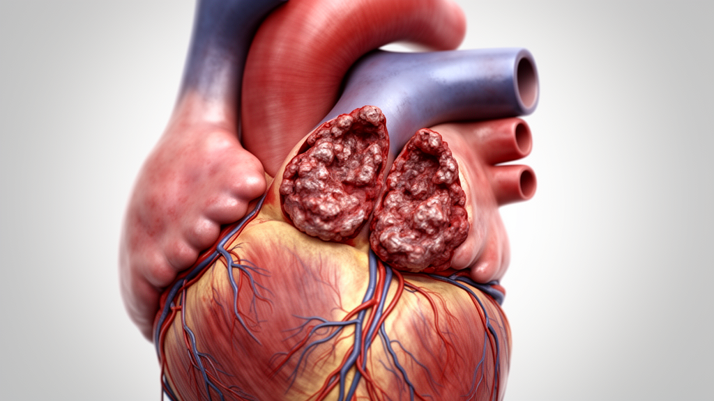
During the surgery, the previously implanted right coronary artery stent was removed due to concerns about persistent infection, and coronary artery bypass grafting to the right coronary artery was added to the procedure. Infected vegetation impacting the ostium of the right coronary artery was directly detected during the operation, confirming coronary embolization as the mechanism of the initial acute coronary occlusion. Although histological examination of the embolic material was not performed, the retrieved material was submitted for microbiological analysis, which yielded methicillin-resistant coagulase-negative Staphylococcus. Blood cultures obtained on admission were also positive for this organism, supporting the diagnosis of septic coronary embolization.
Systemic embolization is a well-known complication of infective endocarditis, yet coronary artery embolism remains rare and is often underdiagnosed. Large cohort studies indicate that embolic events predominantly involve the cerebral and splenic circulations, whereas coronary embolization comprises only a small proportion of cases, accounting for less than 1% to 3% of embolic complications. When coronary embolism does occur, it often manifests as acute coronary syndrome, making the timely differentiation from atherosclerotic plaque rupture or acute coronary thrombosis particularly challenging for clinicians.
Prosthetic valve endocarditis, particularly in the early postoperative period, is associated with aggressive local infection, annular destruction, and poor outcomes. Early prosthetic valve endocarditis can progress rapidly and may present with atypical manifestations before classical signs of infection become apparent. Coronary embolization from vegetations originating on infected prosthetic valves is particularly rare and has been reported only sporadically in the medical literature. In this specific case, the definitive diagnosis was confirmed only after surgical exploration, underscoring the limitations of non-invasive imaging in distinguishing infected vegetation from thrombus.
The diagnostic ambiguity in this case mirrors other reported instances where intravascular ultrasound and contrast-enhanced computed tomography findings were initially interpreted as thrombus, making the initial diagnosis of acute coronary thrombosis reasonable. Coronary embolism caused by septic vegetations presents a substantial diagnostic challenge because its clinical and angiographic features closely resemble those of acute coronary thrombosis. This similarity can lead to delays in recognizing the underlying infectious source, which is critical for effective management. The absence of postoperative anticoagulation following bioprosthetic valve implantation may have also contributed to the initial suspicion of prosthetic valve thrombosis, further complicating the differential diagnosis.
Management of prosthetic valve endocarditis complicated by annular abscess requires radical debridement of infected tissue and secure reconstruction. Extensive annular destruction often necessitates aggressive surgical strategies beyond isolated redo valve replacement. In the present case, near-circumferential annular involvement precluded secure patch reconstruction alone, and aortic root replacement was required to achieve adequate infection control and structural stability. The direct intraoperative visualization of vegetation impacted at the coronary ostium provided unequivocal confirmation that septic coronary embolization was the primary mechanism of the initial clinical presentation.
Despite aggressive surgical and perioperative management, the patient’s condition continued to deteriorate. After successful weaning from extracorporeal membrane oxygenation on postoperative day 6, the patient initially showed transient hemodynamic stabilization. However, he subsequently developed progressive hypotension accompanied by abdominal distension and bloody output from the nasogastric tube, raising strong suspicion of intestinal ischemia. Contrast-enhanced computed tomography revealed no evidence of major mesenteric arterial occlusion; however, extensive ischemic changes were observed in the small intestine, gallbladder, and spleen.
Laboratory findings demonstrated a rapid deterioration, including a marked increase in liver enzymes and lactate dehydrogenase, along with a significant elevation in lactate levels, indicating severe systemic hypoperfusion. Based on these clinical, imaging, and laboratory findings, a diagnosis of non-occlusive mesenteric ischemia was made. Given the patient’s critically unstable hemodynamic condition, the medical team determined that repeat exploratory laparotomy would not provide meaningful therapeutic benefit and would carry a prohibitive risk. Despite intensive supportive care, the patient’s condition progressively worsened, and he died on postoperative day 10.
This case illustrates the systemic severity of early prosthetic valve endocarditis and underscores the limitations of even prompt and radical surgical intervention once profound circulatory collapse has occurred. The complication of non-occlusive mesenteric ischemia was considered a consequence of the prolonged low-output state and severe circulatory failure rather than direct embolic phenomena. The case highlights the importance of maintaining a high index of suspicion for infective endocarditis, even in the absence of definitive early signs of infection.
In conclusion, this case demonstrates that septic coronary embolism should be considered in the differential diagnosis of acute coronary syndrome occurring early after valve surgery. Differentiation between thrombus and vegetation may be extremely difficult using coronary imaging alone. Early valve-focused evaluation and multidisciplinary decision-making are essential to avoid delayed diagnosis in this rare but catastrophic clinical scenario. The findings emphasize the need for careful diagnostic assessment and appropriate postoperative management in patients presenting with acute coronary events following valve replacement.
Subject of Research: Early prosthetic valve endocarditis presenting as acute coronary syndrome
Article Title: Early prosthetic valve endocarditis presenting as acute right coronary artery occlusion 1 month after aortic valve replacement: a case report
Article References: Yokawa, K., Yoshida, K., Ishimoto, K., Nakagawa, T., Tanaka, Y., Higuma, T., Oshima, Y., Obo, H., & Wakiyama, H. (2026). Early prosthetic valve endocarditis presenting as acute right coronary artery occlusion 1 month after aortic valve replacement: a case report. General Thoracic and Cardiovascular Surgery Cases, 5(1), Article 18. https://doi.org/10.1186/s44215-026-00257-2
Image Credits: AI Generated
DOI: 10.1186/s44215-026-00257-2
Keywords: Prosthetic valve endocarditis, Acute coronary syndrome, Coronary embolism, Aortic valve replacement, Cardiogenic shock, Methicillin-resistant Staphylococcus, Non-occlusive mesenteric ischemia, Percutaneous coronary intervention, Early, prosthetic, valve, endocarditis
News Source: Kristina Jarvis. (November 9, 2026). Valve Infection Mimics Heart Attack in Rare Case. Scienmag.