Endovascular aortic repair has established itself as the primary therapeutic modality for abdominal aortic aneurysms, yet type II endoleaks persist as the most prevalent post-procedural complication encountered in clinical practice. A comprehensive narrative review published in CVIR Endovascular synthesizes contemporary evidence on the natural history, diagnostic evaluation, and interventional strategies for these retrogradely perfused vascular cavities, highlighting an essential conceptual shift from technique-centered execution to anatomy-driven therapeutic decision-making. Researchers led by Maciej Błaszyk analyzed medical literature from January 2000 through July 2026 to assess the comparative roles of aneurysm sac and feeding vessel embolization. Their synthesis underscores that procedural success and durable cavity exclusion are primarily governed by vascular architectural complexity and internal flow dynamics rather than by the choice of access route or embolization tool alone.
In post-repair pathology, endoleaks are formally categorized into five discrete anatomical classifications, with persistent blood flow occurring in approximately 10 to 40 percent of endovascular aneurysm repair interventions. Type II endoleaks represent retrograde perfusion of the excluded aneurysm sac via branching collateral vessels and account for roughly half of all diagnosed endoleaks. While attachment-site disruptions, structural graft defects, graft fabric porosity, and endotension constitute other endoleak varieties, the natural progression of isolated branch-fed leaks is predominantly benign. Up to one-third to one-half of these occurrences resolve spontaneously without any invasive intervention, and observational investigations have repeatedly noted that the risk of catastrophic aneurysm rupture remains below 1 percent, even though rupture events across all abdominal aortic aneurysm etiologies carry an overall mortality rate approximating 81 percent.
Clinical surveillance guidelines delineate strict thresholds before invasive reintervention is warranted for a persistent type II endoleak. Both the 2024 Cardiovascular and Interventional Radiological Society of Europe standards and the 2024 European Society of Vascular Surgery clinical practice guidelines advise conservative watchful waiting for stable aneurysmal sacs. Secondary interventional therapy is formally recommended only after the definitive exclusion of high-flow type I attachment leaks or type III graft integrity defects, and specifically when persistent retrograde perfusion is accompanied by significant sac expansion of at least 10 millimeters compared with baseline or the smallest documented diameter during follow-up, or in rare cases of related complications such as sac rupture.
The underlying hemodynamic pathophysiology of these endoleaks is driven by delicate pressure gradients established between the excluded aneurysm cavity and originating arterial branches. Experimental models demonstrate that relatively elevated pressure within the inferior mesenteric artery frequently positions it as a dominant inflow channel. Morphologically, these leaks are divided into type IIa patterns, which feature a single feeding vessel, and type IIb configurations, which involve multiple interconnected collateral vessels that establish reciprocal inflow and outflow channels coursing directly through the sac cavity. While single-vessel type IIa configurations demonstrate a higher propensity for spontaneous resolution, the interconnected circuits of type IIb endoleaks are far more likely to persist and induce progressive aneurysmal enlargement.
Diagnostic and procedural imaging plays a vital role in determining whether therapeutic intervention is indicated and in defining the precise anatomical target. Computed tomography angiography remains the primary diagnostic reference standard for longitudinal post-repair surveillance, utilizing standardized imaging at 30 days and extended intervals to monitor sac behavior. However, standard imaging frequently struggles to differentiate between simple and complex inflow-outflow pathways. Dynamic techniques such as time-resolved computed tomography angiography, four-dimensional flow-sensitive magnetic resonance imaging, and contrast-enhanced ultrasound provide superior real-time characterization of flow velocity and directional hemodynamics. Contrast-enhanced ultrasound offers sensitivity comparable to computed tomography without exposing patients to ionizing radiation, though its diagnostic accuracy can be compromised by excessive bowel gas, severe patient obesity, and operator dependency.
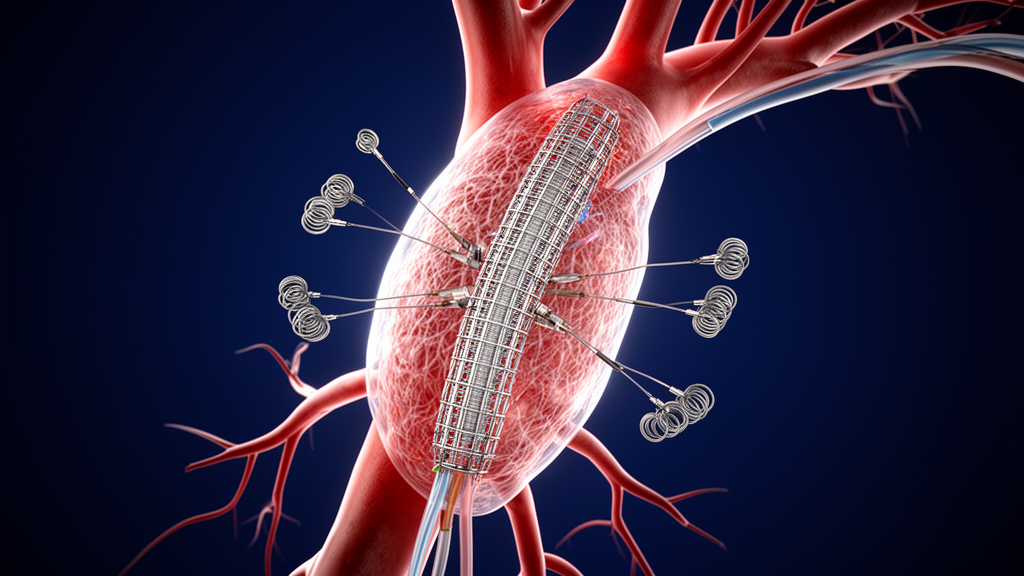
When intervention becomes mandatory due to progressive sac expansion, interventional radiologists choose between targeting the feeding branches, the interior aneurysm cavity, or both vascular zones simultaneously. Transarterial embolization remains the most commonly deployed first-line technique due to its minimally invasive transfemoral or transbrachial access routes and broad operator familiarity. In simple type IIa cases involving an accessible dominant vessel, such as a large-caliber inferior mesenteric artery or a reachable lumbar branch, selective transarterial catheterization can successfully occlude the inflow channel. However, when complex collaterals are present, isolated feeder occlusion frequently proves insufficient because persistent retrograde blood flow readily continues through adjacent communicative pathways, resulting in clinical failure and recurrent sac pressurization.
To mitigate persistent perfusion in anatomically intricate circuits, sac-directed strategies aim to completely disrupt the central hemodynamic nidus. Direct sac puncture, performed percutaneously via transabdominal or translumbar approaches under fluoroscopic, ultrasound, or computed tomography guidance, enables operators to deliver embolic agents directly into the active cavity. Comparative clinical literature indicates that direct sac puncture achieves technical success rates reaching up to 97 percent and clinical success rates ranging between 78 and 81 percent, whereas transarterial techniques report technical feasibility between 71 and 87 percent alongside widely variable clinical success. Furthermore, procedural timing analyses reveal that direct sac puncture entails significantly shorter mean fluoroscopy durations and reduced overall procedure lengths compared with protracted transarterial catheterizations through tortuous collateral networks.
The physical composition of selected embolic materials also contributes significantly to procedural durability, encompassing metallic coils and rapidly precipitating liquid embolic agents. Metallic coils are predominantly deployed for mechanical occlusion of larger side branches; however, monotherapy with coils alone has been linked to higher rates of reintervention compared with liquid agents. Liquid embolics include ethylene-vinyl alcohol copolymer, which precipitates into a spongy, cohesive cast as its dimethyl sulfoxide solvent disperses into surrounding blood, and N-butyl cyanoacrylate, a monomer that polymerizes rapidly upon contact with ionic blood components. Liquid agents possess the distinct rheological capacity to migrate through irregular nidus channels and seal hidden outflow channels without requiring superselective catheter engagement of every individual collateral vessel, though ethylene-vinyl alcohol copolymer entails considerable acquisition expense and lacks defined maximum safe volumetric thresholds.
Despite apparent technical advantages and shorter procedural durations associated with direct percutaneous sac access, investigators emphasize that direct comparisons across published studies are heavily confounded by methodological heterogeneity. Published series differ extensively in outcome definitions, defining clinical success inconsistently as the complete absence of flow, the cessation of sac enlargement, or explicit dimensional sac shrinkage. Moreover, direct sac puncture is disproportionately reserved for refractory failures and highly convoluted anatomical presentations, introducing unavoidable patient selection bias. As a result, current medical evidence does not substantiate the absolute superiority of one universal interventional modality over another, nor does randomized data support pre-emptive prophylactic feeder embolization prior to the index aortic repair.
Consequently, the authors propose a practical clinical decision framework that stratifies interventions according to specific vascular architecture and flow complexity. Anatomically straightforward endoleaks with an accessible dominant channel and limited collaterals can be managed effectively with selective transarterial feeder and cavity occlusion. Conversely, complex multi-branch type IIb circuits, ambiguous inflow-outflow pathways, and instances of failed prior transarterial attempts necessitate sac-centered eradication via direct percutaneous puncture, translumbar access, or alternative adjunctive techniques such as transiliac paraendograft or transcaval approaches. By shifting procedural planning away from rigid technical habits and toward an individualized, anatomy-driven evaluation of the hemodynamic circuit, clinical teams can optimize procedural safety and achieve durable exclusion for expanding aneurysm sacs.
Beyond vascular morphology, the review highlights several patient-level risk indicators and systemic influences that govern endoleak behavior. Advanced age, larger initial aneurysm diameter, a patent inferior mesenteric artery, and a higher count of patent lumbar arteries correlate with higher rates of occurrence and persistence. Conversely, the presence of fewer than six patent lumbar arteries is associated with a greater likelihood of spontaneous sac reduction. Risk stratification models, such as the classification proposed by Mo and colleagues, combine lumbar vessel counts with mesenteric arterial caliber to categorize patients into low-, intermediate-, and high-risk profiles for targeted surveillance. Counterintuitively, active smoking and chronic obstructive pulmonary disease show an inverse association with endoleak formation. The authors note that this apparent protective effect is likely driven by smoking-induced vascular alterations and accelerated accumulation of intraluminal thrombus within the aneurysm sac, which physically occludes potential feeding branch orifices and prevents retrograde filling.
Procedural timing and intraoperative imaging also present notable operational differences across intervention types. While direct percutaneous sac puncture requires careful navigation around the inferior epigastric artery, mesenteric vessels, and bowel—maintaining constant needle pressure to displace intestinal loops—it demonstrates distinct time efficiencies. In comparative analyses by Yang and colleagues, direct sac puncture achieved significantly shorter mean fluoroscopy times of 11.2 minutes compared with 42.4 minutes for transarterial approaches, alongside shorter overall procedure durations of 103 minutes versus 181 minutes. Independent series by Zener and Fanelli recorded median or mean fluoroscopy durations ranging from 11.3 to 16.7 minutes. The authors emphasize, however, that fluoroscopy duration is not equivalent to cumulative radiation exposure. To enhance procedural precision, clinicians increasingly utilize intraoperative cone-beam computed tomography, which assists catheter navigation and puncture targeting while potentially reducing contrast-agent administration.
For anatomical configurations where standard access routes are unavailable, the review outlines specialized adjunctive techniques. These include entering the aneurysm cavity through the potential space between the arterial wall and graft limb via a transiliac paraendograft pathway, or performing transcaval puncture through the inferior vena cava, which proves advantageous when the endoleak nidus is situated on the right side of the sac. In select direct puncture cases, agents such as thrombin or gelatin-based materials provide alternative embolization options. When all endovascular and percutaneous attempts fail, open surgical repair remains the definitive treatment option, achieving technical success rates approaching 100 percent and durable sac stabilization at the expense of substantial surgical invasiveness.
Subject of Research: Contemporary embolization strategies and anatomy-driven decision-making for type II endoleaks following endovascular aortic repair
Article Title: Targeting the sac or the feeders? An anatomy-driven review of contemporary embolization strategies of type II endoleaks
Article References: Błaszyk, M., Waliszewski, J., Fryska, Z., Gargula, D., Wruck, P., Karbowiak, K., & Juszkat, R. (2026). Targeting the sac or the feeders? An anatomy-driven review of contemporary embolization strategies of type II endoleaks. CVIR Endovascular, 9(1), Article 106. https://doi.org/10.1186/s42155-026-00761-0
Image Credits: AI Generated
DOI: 10.1186/s42155-026-00761-0
Keywords: Endovascular Aortic Repair, Type II Endoleaks, Embolization Strategies, Direct Sac Puncture, Transarterial Embolization, Aneurysm Sac Enlargement, Vascular Anatomy, Liquid Embolics, Interventional Radiology, Computed Tomography Angiography, Targeting, feeders
News Source: Ophelia Keating. (November 9, 2026). Anatomy-Driven Approaches Guide Embolization Decisions for Post-EVAR Type II Endoleaks. Scienmag.