
Thanatophoric dysplasia (TD) is an intractable disease designated by Ministry of Welfare, Health and Labor (MWHL) of Japan, causing severe abnormalities of bones and the brain. The limb and rib bones are shorter than normal ones, and as regards the brain, abnormalities such as polymicrogyria*1 and periventricular nodular heterotopia (PNH)*2 are discerned. As a gene responsible for TD, fibroblast growth factor receptor 3 (FGFR3) has been reported. However, since it is a rare disease and since it is difficult to obtain brain samples from human patients, the pathophysiology of TD is largely unknown, and effective therapy has not been established.
Concerning the bone abnormalities in TD patients, a team of Kyoto University reported the research results using iPS cells (Nature 2014, 513, 507-511), which attracted much attention. On the other hand, research on abnormalities of the brain has been rather retarded.
The present research team of Kanazawa University succeeded in generating an animal model using ferrets*3, which reproduces the brain abnormalities found in human TD patients. By using this animal model, the team elucidated the formation process of polymicrogyria, one of the abnormalities found in the TD brain. In the present study, the team has investigated the formation process of PNH, the other brain abnormality found in human TD patients, using their animal model of TD.
[Results]
The present research team has succeeded in uncovering the pathophysiological process of the formation of PNH by using the TD ferrets as follows.
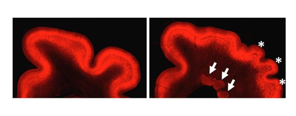
First, PNH was analyzed in terms of composing cell types to reveal that a large number of neurons but few glial cells*4 existed in PNH. In healthy brain, neurons are found in the cerebral cortex near the brain surface. It was, therefore, considered that during the fetal brain development, PNH formation might be induced by inability of neurons to translocate themselves to their destination, that is, the cerebral cortex. Next, the reason why neurons were unable to translocate themselves was investigated. It was found that spatial arrangement of radial glial cells*5 were distorted; radial glial fibers were considered to serve as the track for neurons to translocate themselves. Thus, it was suggested that the distortion of radial glial fibers would be a reason for aberrant localization of neurons.
[Significance]
The research team elucidated the important process of the PNH formation mechanism. It is only possible, by using appropriate animal model that reproduces relevant pathophysiology, to uncover the process of pathogenesis and to develop therapy. Since the research on abnormalities of bones in TD is progressing with iPS cells at Kyoto University, it is expected that the whole aspect of TD with brain and bone abnormalities would be elucidated and that the therapeutic methods would be developed.
The research team has recognized the importance of disease model animals with the brain architecture more similar to that of humans than mice, a typical model animal, and has been uniquely in the world developing experimental techniques for ferrets, a higher mammal, of Mustelidae. The present study on PNH was only possible with the experimental technique for ferrets developed by the research team. It is expected with the unique experimental technique that research will be carried out further on neurological diseases that have been difficult to investigate with conventional model animal, mouse, and that therapy will be developed in a more accelerated manner.
###
[Glossary]
*1Polymicrogyria
The surface of the cerebrum has many folds called the gyrus. In the patients of thanatophoric dysplasia (TD), abnormalities of the gyri are seen. More specifically, a larger number of smaller gyri are seen than normal. Hence it is called polymicrogyria.
*2Periventricular nodular heterotopia (PNH)
One of the abnormalities seen in the TD brain is PNH. In normal brain, neurons exist in the cerebral cortex whereas in the brain with TD, some neurons exist in the deep part of the brain as clusters, which are called PNH. The mechanism of PNH formation was largely unknown.
*3Ferret
A higher mammal of Mustelidae. Since ferrets have the brain more developed than mice and rather similar to the human brain, ferrets were used as model animal in this study. Genetic research using ferrets are not yet widely carried out in the world, and the present research team is unique in this regard.
*4Glial cells
Most of cells other than neurons in the brain are called glial cells. Glial cells are thought to be important in various functions to support neurons.
*5Radial glia
Radial glia are special cells seen during the period of fetus brain development. They are also referred to as neural stem cells, which develop into neurons and glial cells. In addition, their fibers serve as the track for neurons generated deep in the brain to translocate themselves toward the brain surface.
Media Contact
Takashi Shimizu
[email protected]
81-762-645-963
http://www.kanazawa-u.ac.jp/e/index.html
