Researchers have demonstrated that noninvasive oxygen-enhanced brain magnetic resonance imaging combining dynamic three-dimensional gradient-echo imaging and three-dimensional magnetic resonance fingerprinting can visualize and quantitatively track the transit of inhaled oxygen into surface cerebrospinal fluid. In a prospective study evaluating twenty healthy participants enrolled between December 2023 and March 2024, dynamic volumetric interpolated breath-hold examination revealed immediate signal increases across the subarachnoid cerebrospinal fluid space surrounding the cerebral cortex following supplemental oxygen inhalation, with secondary signal changes detected within cerebral gray matter. These dynamic signal alterations were substantiated through quantitative longitudinal relaxation time reductions derived from expanded three-dimensional magnetic resonance fingerprinting dictionaries, confirming physiological oxygen transit into fluid compartments adjacent to brain parenchyma.
Understanding cerebrospinal fluid dynamics has gained heightened scientific interest alongside investigations into the glymphatic system, a pathway through which cerebrospinal fluid permeates the brain parenchyma while bypassing the blood-brain barrier to facilitate solute and interstitial fluid exchange toward perivenous spaces. Previous clinical investigations into cerebrospinal fluid dynamics frequently depended on the intrathecal administration of gadolinium-based contrast agents, which exhibited centripetal parenchymal enhancement over hours and cleared over several weeks. However, intrathecal gadolinium administration faces widespread regulatory restrictions because of associated health risks. Other noninvasive approaches have explored indirect kinetic imaging with oxygen-17 labeled water, while oxygen-enhanced magnetic resonance imaging takes advantage of the natural longitudinal relaxation shortening effect induced by dissolved molecular oxygen during hyperoxic inhalation.
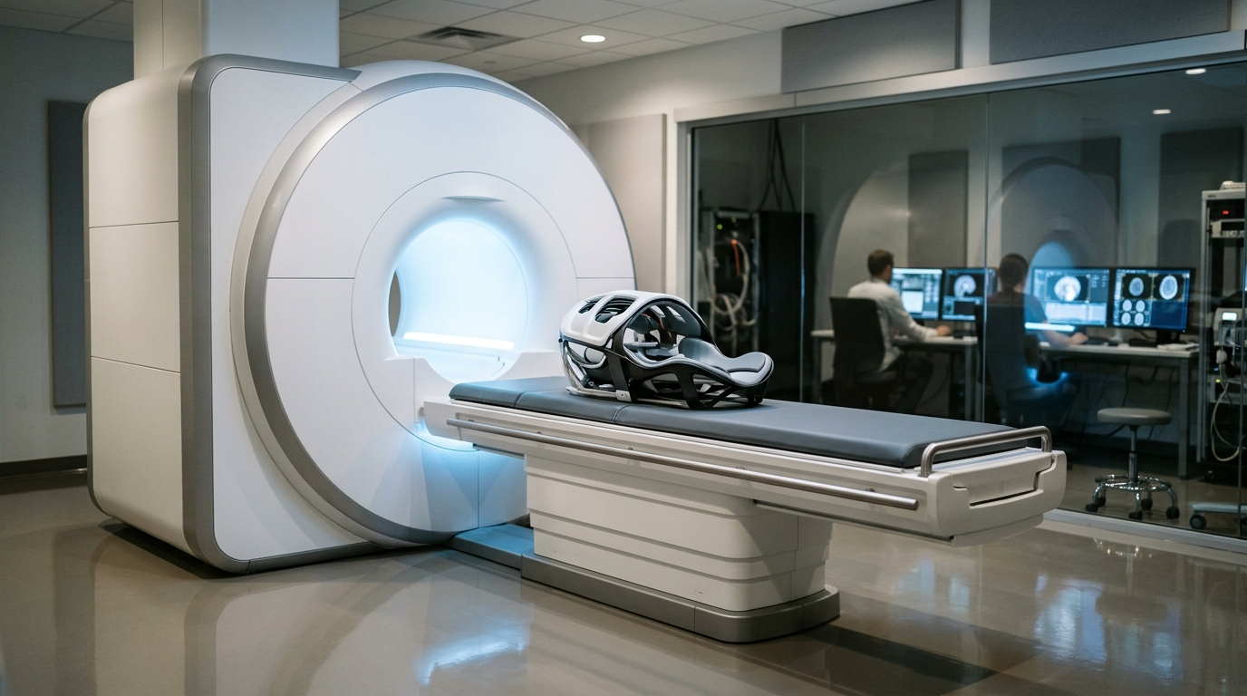
The experimental protocol was conducted on a 3-Tesla Siemens Magnetom Prisma scanner equipped with a 20-channel receive-only head and neck coil. The cohort consisted of twenty healthy volunteers with a mean age of 74.4 plus or minus 4.8 years, comprising 11 females and 9 males, who completed the entire imaging procedure without adverse events or complications. Participants were administered supplemental oxygen via a reservoir mask from Intersurgical. Prior to hyperoxic challenge, participants breathed ambient room air through air vents supplemented with a minimal baseline flow of 1 liter per minute of oxygen to prevent carbon dioxide rebreathing inside the mask structure. Full hyperoxic administration was initiated at the eighth dynamic measurement using a high-flow delivery rate of 15 liters per minute, which was continuously sustained throughout subsequent quantitative imaging runs.
Dynamic imaging utilized a three-dimensional volumetric interpolated breath-hold examination gradient-echo sequence acquired across 20 serial repetitions, requiring 15 seconds per measurement for a total acquisition duration of 5 minutes. The sequence operated with a repetition time of 4.25 milliseconds, an echo time of 1.67 milliseconds, a flip angle of 15 degrees, an isotropic voxel size of 2 by 2 by 2 millimeters cubed, a field of view of 256 by 256 millimeters squared, a matrix resolution of 128 by 128, and GRAPPA parallel imaging acceleration of 2 by 2. To eliminate initial signal instability analogous to functional neuroimaging processing, the first volumetric measurement was excluded. Dynamic ratio curves were calculated by dividing signal intensities at each time frame by the second measurement, normalizing pre-inhalation baseline values across measurements three through seven to unity.
Quantitative mapping relied on three-dimensional magnetic resonance fingerprinting performed before and after oxygen administration, designated as Pre-1, Pre-2, Post-1, and Post-2. Each magnetic resonance fingerprinting run lasted 5 minutes and 46 seconds, employing multiaxis variable-density spiral projection k-space trajectories with zero-moment nulling to capture an isotropic spatial resolution of 1 by 1 by 1 millimeter cubed across a 240 by 240 by 240 millimeters cubed field of view. To accommodate the prolonged relaxation rates of pure fluid, the precalculated dictionary was extended to cover longitudinal relaxation times from 0 to 5,000 milliseconds. Across 30 acquisition blocks containing 500 repetition times each, sequence preparation used adiabatic inversion pulses with an inversion time of 20 milliseconds, sinusoidal flip angles ranging from 5 to 82 degrees, repetition times of 16 milliseconds, and echo times of 2.5 milliseconds.
Image reconstruction and post-processing incorporated sliding-window data combinations to fill spherical k-space, three-dimensional inverse nonuniform fast Fourier transforms, and ESPIRiT coil sensitivity estimation on an Ubuntu workstation running MATLAB R2019b. Anatomical structural reference images were acquired using three-dimensional magnetization-prepared rapid gradient-echo sequences with isotropic 0.9-millimeter voxels accelerated by WaveCaipi 6-fold undersampling. Brain segmentation masks established three-dimensional volumes of interest for gray matter, white matter, surface cerebrospinal fluid, and ventricular compartments at a probability threshold of 0.3. For group comparisons, volumes were spatially normalized to Montreal Neurological Institute coordinate space using DARTEL diffeomorphic anatomical registration algorithms, while native space volumes were additionally evaluated using one-voxel eroded margins to minimize partial volume averaging.
Statistical evaluation using paired Wilcoxon signed-rank tests with false discovery rate correction demonstrated that post-inhalation volumetric interpolated breath-hold examination signals were significantly elevated compared to pre-inhalation levels in surface cerebrospinal fluid and gray matter, both achieving significance thresholds below 0.001. Signal elevation in surface cerebrospinal fluid displayed a very large within-subject effect size, quantified by a Cohen dz of 4.15, while gray matter exhibited an effect size of 1.31. In contrast, white matter and ventricular spaces showed negligible effect sizes of 0.01 and 0.14, respectively. Regression slopes across post-inhalation intervals confirmed that the surface cerebrospinal fluid space exhibited the steepest and most significant rate of signal increase among all evaluated regions, while pre-inhalation baseline coefficients of variation confirmed minimal drift.
Quantitative longitudinal relaxation measurements from magnetic resonance fingerprinting revealed significant phase-dependent variations across all evaluated compartments, showing omnibus Friedman test values of p less than 0.001 for gray matter, white matter, and surface cerebrospinal fluid, alongside p equal to 0.024 for ventricles. In surface cerebrospinal fluid, mean longitudinal relaxation times decreased from a Pre-1 baseline of 2,512.4 milliseconds and Pre-2 baseline of 2,505.9 milliseconds down to 2,332.7 milliseconds at Post-1 and 2,351.3 milliseconds at Post-2, reflecting a within-subject effect size of Cohen f equal to 1.31. Post hoc pairwise tests via Conover comparisons confirmed that Post-1 and Post-2 fluid relaxation times were significantly lower than pre-oxygen levels (p less than 0.001). Gray matter relaxation similarly dropped from a Pre-1 mean of 1,516.7 milliseconds to 1,474.7 milliseconds at Post-1 (p less than 0.001, Cohen f equal to 0.73).
Supplementary assessments confirmed the stability of these physiological findings across independent secondary metrics. Assessments using one-voxel eroded masks corroborated significant signal and relaxation shifts for gray matter and surface cerebrospinal fluid (p less than 0.001), indicating that findings were not driven solely by border partial volume effects. Test-retest reliability across pre-inhalation baselines demonstrated two-way random-effects intraclass correlation coefficients of 0.963 for surface cerebrospinal fluid, 0.917 for white matter, 0.825 for gray matter, and 0.714 for ventricular spaces. Supplemental longitudinal rate calculations and relaxation time ratios relative to Pre-1 verified matching dynamic phase distinctions across tissue types, while Cramer-Rao lower bound analyses indicated high precision across parenchymal regimes despite reduced estimation sensitivity for extremely long fluid relaxation components.
The study authors emphasize several critical technical and physiological limitations regarding the interpretation of these oxygen-enhanced magnetic resonance observations. While dissolved paramagnetic oxygen induces observable shortening of longitudinal relaxation, absolute signal increases on gradient-echo images remained modest at approximately 2 to 3 percent, and true bolus delivery could not be achieved because gas intake depended on spontaneous respiration. Furthermore, the findings reflect oxygen transit into subarachnoid fluid adjacent to cortical arterial vasculature rather than direct proof of parenchymal interstitial clearance or glymphatic flushing. The authors also noted that participants were older healthy volunteers with high health literacy, physiological variables such as arterial oxygen saturation and end-tidal carbon dioxide were unmonitored, transverse relaxation times were not acquired, and unmeasured hyperoxic vasoconstriction or mask-related carbon dioxide accumulation could have influenced regional cerebral blood flow.
Subject of Research: Oxygen-enhanced magnetic resonance imaging of human cerebrospinal fluid dynamics
Article Title: Oxygen-enhanced brain MRI using dynamic gradient-echo and MR fingerprinting
Article References: Fushimi, Y., Okuchi, S., Utsunomiya, Y., Nakajima, S., Sakata, A., Yamamoto, T., Oshima, S., Liao, C., Imai, H., & Nakamoto, Y. (2026). Oxygen-enhanced brain MRI using dynamic gradient-echo and MR fingerprinting. European Radiology Experimental, 10(1), Article 174. https://doi.org/10.1186/s41747-026-00836-6
Image Credits: AI Generated
DOI: 10.1186/s41747-026-00836-6
Keywords: Magnetic Resonance Imaging, Cerebrospinal Fluid, MR Fingerprinting, Oxygen-Enhanced MRI, Brain Imaging, Neuroimaging, Glymphatic System, VIBE Sequence, Oxygen-enhanced, brain, dynamic, gradient-echo
News Source: Cassandra Pierce. (November 7, 2026). Dynamic MRI Protocol Tracks Oxygen Movement Across Human Brain Fluid Compartments. Scienmag.