A synchronized investigation of visual target acquisition, cortical electrical activity, and peripheral muscle recruitment has revealed that subacute stroke survivors preserve a measurable temporal window of motor preparation despite extensive delays across the entire sensorimotor cascade. Investigating how upper-limb impairment alters the fine-grained timing between intention and movement, researchers evaluated thirty subacute stroke patients alongside thirty demographically matched healthy control participants during a standardized full-plane reaching task. By simultaneously acquiring high-density eye-tracking data, 64-channel electroencephalography, and multi-channel surface electromyography, the study established the first trial-level interval structure charting when successive physiological stages become active within the same individual. The experimental results demonstrated that visual attention engagement and peripheral muscular contraction do not merely lag behind normative schedules in isolation; rather, they exhibit a coordinated, stepwise temporal delay that brackets a distinct epoch of preparatory brain activity. These findings provide an empirical, neurophysiological baseline that describes how subcortical stroke reconfigures the chronological sequence linking sight, cortical readiness, and physical reach.
Upper-limb hemiparesis remains one of the most prevalent and debilitating consequences of stroke, affecting approximately eighty percent of survivors and severely restricting their autonomy in everyday activities. While robotic exoskeletons and functional electrical stimulation offer therapeutic potential during the critical subacute recovery window, their therapeutic efficacy has long been presumed to rely upon the precise temporal synchronization of external assistance with residual voluntary effort. Conventional interventions frequently administer continuous stimulation or depend on therapist-triggered cues, yet meta-analytic reviews demonstrate that assistive technologies gated on physiological states provide primarily modest, heterogeneous functional gains. A persistent engineering bottleneck has been the absence of precise trial-level measurements detailing the chronological sequence of motor planning stages within an individual participant. Previous investigations typically evaluated single physiological channels independently, relying solely on electromyography to register ongoing muscular execution or electroencephalography to decode motor intentions from damaged cortical networks. Because peripheral sensors cannot register events prior to physical movement initiation, and damaged cortices yield variable planning signals, identifying the exact lead times separating ocular engagement, motor cortex commitment, and muscle onset has remained an unresolved clinical priority.
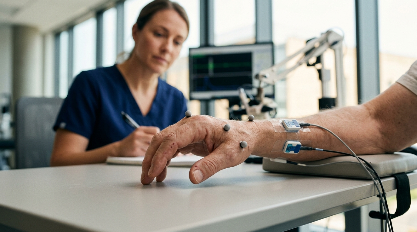
To rigorously map this timing sequence under uniform spatial and biomechanical demands, the investigators implemented an event-related reaching protocol across a full-plane nine-grid display. The visual interface presented a three-by-three matrix of equidistant circular targets arranged to prevent spatial position from introducing systematic biases into search durations or electromechanical responses. Each participant completed alternating blocks consisting of nine pseudo-randomized target displays shown for 4,000 milliseconds, separated by central fixation crosses presented for 2,000 milliseconds, while executing forward reaches with each arm. Eye movements were captured continuously using a Tobii Pro Spectrum tracker operating at 1,200 Hertz, while electroencephalographic potentials were recorded via a Neuroscan SynAmp2 amplifier at 1,000 Hertz, and electromyographic signals were acquired bilaterally from five upper-limb muscles at 3,000 Hertz. To ensure temporal fidelity and analytical alignment across diverse physiological sensors, all three data streams were downsampled to a shared 600 Hertz framework. The cohort consisted of subacute patients with first-ever unilateral supratentorial lesions strictly confined to subcortical structures, evaluated at a median interval of one month following stroke onset, alongside healthy volunteers presenting comparable age, sex, and educational distributions.
Analysis of ocular metrics demonstrated that subacute stroke profoundly alters visual exploration and target acquisition behavior prior to limb movement. When targets appeared on the display matrix, stroke survivors exhibited a significant delay in directing their visual attention toward the designated area of interest, registering a mean time to first fixation of 616.73 milliseconds compared to 367.86 milliseconds in healthy controls. This substantial ocular latency difference was accompanied by marked shifts in gaze structure: stroke patients displayed significantly diminished average fixation durations across the trial, reduced overall fixation density, and a broader spatial spread of gaze points across the workspace. While healthy individuals rapidly localized the visual objective and maintained steady foveal contact, patients exhibited prolonged search patterns and fragmented fixation dynamics. The authors noted that this visual engagement latency carries direct implications for rehabilitation technology design, which frequently employs brief visual prompts calibrated to healthy performance standards. Instead, subacute stroke patients required more than 600 milliseconds of dedicated visual engagement before the target could be fully processed to guide downstream motor planning.
Cortical electrophysiology recorded over bilateral primary sensorimotor regions underscored that the delay in visual engagement propagated directly into the early stages of cortical motor planning. Power spectral density analyses revealed that stroke patients exhibited significantly attenuated relative Beta-band power over the C3 and C4 electrodes during movement preparation compared to healthy subjects, alongside elevated relative Theta-band activity over C4. Because Beta-band desynchronization serves as a primary electrophysiological signature of cortical motor activation and readiness, diminished Beta power reflects compromised planning capacity within surviving motor networks. To determine the exact onset of lateralized motor commitment, the authors derived stimulus-locked lateralized readiness potentials by computing contralateral-minus-ipsilateral difference waves across hand conditions. In the patient group-average waveform, the lateralized readiness potential crossed the pre-stimulus baseline zero line at 854 milliseconds post-stimulus onset, whereas healthy control difference waves showed no crossing under identical baseline criteria. Extensive sensitivity analyses applying varied amplitude thresholds and baseline intervals confirmed that the patient preparatory onset consistently fell between 833 and 930 milliseconds, demonstrating robust cortical readiness preceding muscular recruitment.
Electromyographic recordings from the clinically affected upper limb demonstrated pronounced neuromuscular delays and compensatory recruitment patterns during reaching execution. The anterior deltoid muscle, which serves as the primary agonist for forward shoulder flexion during the planar reaching task, displayed electromechanical onset at 1,080 milliseconds after stimulus presentation in stroke patients, contrasting sharply with the 605 milliseconds recorded in healthy controls. In addition to this 475-millisecond execution delay, the patient group exhibited exaggerated anterior deltoid integrated electromyography and elevated median frequency, culminating in a mean peak activation amplitude of 143.15 microvolts compared to 84.74 microvolts in healthy subjects. Concurrently, stroke survivors exhibited elevated median frequency in the upper trapezius and reduced root-mean-square amplitude in the biceps brachii, reflecting disorganized multi-muscle synergy and compensatory recruitment. Crucially, the chronological gap between the lateralized readiness potential onset at 854 milliseconds and anterior deltoid contraction at 1,080 milliseconds confirms that a 226-millisecond cortical preparation window survives stroke damage, existing downstream of delayed vision and ahead of abnormal muscular hyperactivation.
Leveraging these temporal milestones, the researchers investigated whether ocular dynamics alone could supply sufficient discriminative information to identify when a patient prepares to execute a goal-directed movement. Segmenting eye-tracking recordings across all sixty participants produced 1,125 valid analysis windows, encompassing 534 target-presentation reaching intervals and 591 inter-target rest periods. Nine normalized, window-level gaze features were extracted from the event streams, including time to first fixation, fixation and saccade rates, saccade amplitude, gaze spread, fixation density, and gaze entropy, each mathematically screened to eliminate artificial correlations with window length. Five distinct machine-learning models were trained and tested using participant-level leave-one-subject-out cross-validation, guaranteeing that no held-out participant contributed to feature scaling, parameter selection, or training. Under this rigorous evaluation, a random forest classifier attained an area under the receiver operating characteristic curve of 0.973, achieving an overall classification accuracy of 0.918, a sensitivity of 0.897, and a specificity of 0.937, outperforming AdaBoost, convolutional-recurrent architectures, and support vector machines.
To evaluate whether this elevated classification performance reflected genuine sensorimotor patterns or subtle artifacts of methodological leakage, the researchers conducted extensive validation and ablation procedures. Permuting class labels forty times across participants reduced the receiver operating characteristic curves to median null values of 0.492 and 0.498, verifying the complete absence of data leakage and validating the independence of the cross-validation structure. Repeated random participant partitions utilizing alternate eight-to-two and two-to-one ratios yielded median area-under-the-curve metrics exceeding 0.97, confirming that classifier stability did not depend on idiosyncratic training splits. Feature importance assessments demonstrated that the latency to first fixate the target area of interest carried the largest proportion of discriminative power; completely removing this single timing feature caused the random forest model area under the curve to drop to 0.871. These analyses confirmed that the classifier successfully exploited the predictable pathophysiological delays that accompany instructed reaching rather than learning static participant characteristics.
A critical secondary finding emerged when the researchers evaluated whether ocular, cortical, or muscular features could differentiate stroke patients from healthy individuals at the group level under identical leave-one-subject-out folds. Classifying participant clinical diagnosis using peripheral electromyographic features achieved an area under the curve of 0.838, whereas cortical spectral features attained an intermediate score of 0.735, and ocular features remained entirely at chance level with an area under the curve of 0.456. Combining all three measurement modalities failed to surpass the predictive accuracy of the peripheral muscle signals alone. This stark dissociation illustrates that while gaze dynamics cannot distinguish a patient from a control, they reliably track within-participant state transitions between rest and motor intention. For practical translation into neurorehabilitative devices, this functional distinction is advantageous: an assistive orthosis or functional electrical stimulation controller does not require biometric user identification, but instead requires an unobtrusive, continuous signal capable of detecting when that specific user transitions into active movement preparation.
Despite these promising classification benchmarks, the investigators emphasized several structural limitations that circumscribe the clinical application of their findings. The patient cohort was evaluated exclusively between one and three months post-stroke, a period characterized by dynamic neuroplasticity and spontaneous biological recovery; consequently, these temporal latencies represent stage-specific values that cannot be extrapolated to chronic populations without dedicated re-calibration. Additionally, the experimental models functioned offline on complete observation windows, lacking real-time, closed-loop execution under active mechanical guidance. Because every participant presented subcortical damage without cortical lesion involvement, the observed sequence cannot be generalized to cortical strokes or varied structural lesion loads without independent verification. The authors caution that their data delineate an empirical, testable physiological framework rather than direct proof of clinical therapeutic benefit. Translating this preserved preparation window into functional motor recovery will require prospective, closed-loop clinical trials that evaluate whether delivering assistance prior to abnormal peripheral muscle recruitment truly enhances long-term neuroplasticity in broader stroke populations.
Subject of Research: Temporal dynamics of the sensorimotor cascade and gaze classification in subacute stroke
Article Title: Temporal dissociation across the eye-brain-muscle cascade delineates a preparation window before muscle activation in subacute stroke
Article References: He, Y., Hu, Y., Wu, X., Zhang, J., Liu, Z., Huang, J., Lu, X., Wu, X., Wu, J., Chen, L., & Tao, J. (2026). Temporal dissociation across the eye-brain-muscle cascade delineates a preparation window before muscle activation in subacute stroke. Frontiers in Human Neuroscience, 20, Article 2002490. https://doi.org/10.3389/fnhum.2026.2002490
Image Credits: AI Generated
DOI: 10.3389/fnhum.2026.2002490
Keywords: stroke rehabilitation, sensorimotor cascade, electroencephalography, surface electromyography, eye tracking, lateralized readiness potential, motor preparation, neurotechnology, Temporal, dissociation, across, eye-brain-muscle
News Source: Cassandra Pierce. (November 7, 2026). Sensorimotor Cascade Timing Uncovers a Neural Preparation Window in Subacute Stroke. Scienmag.