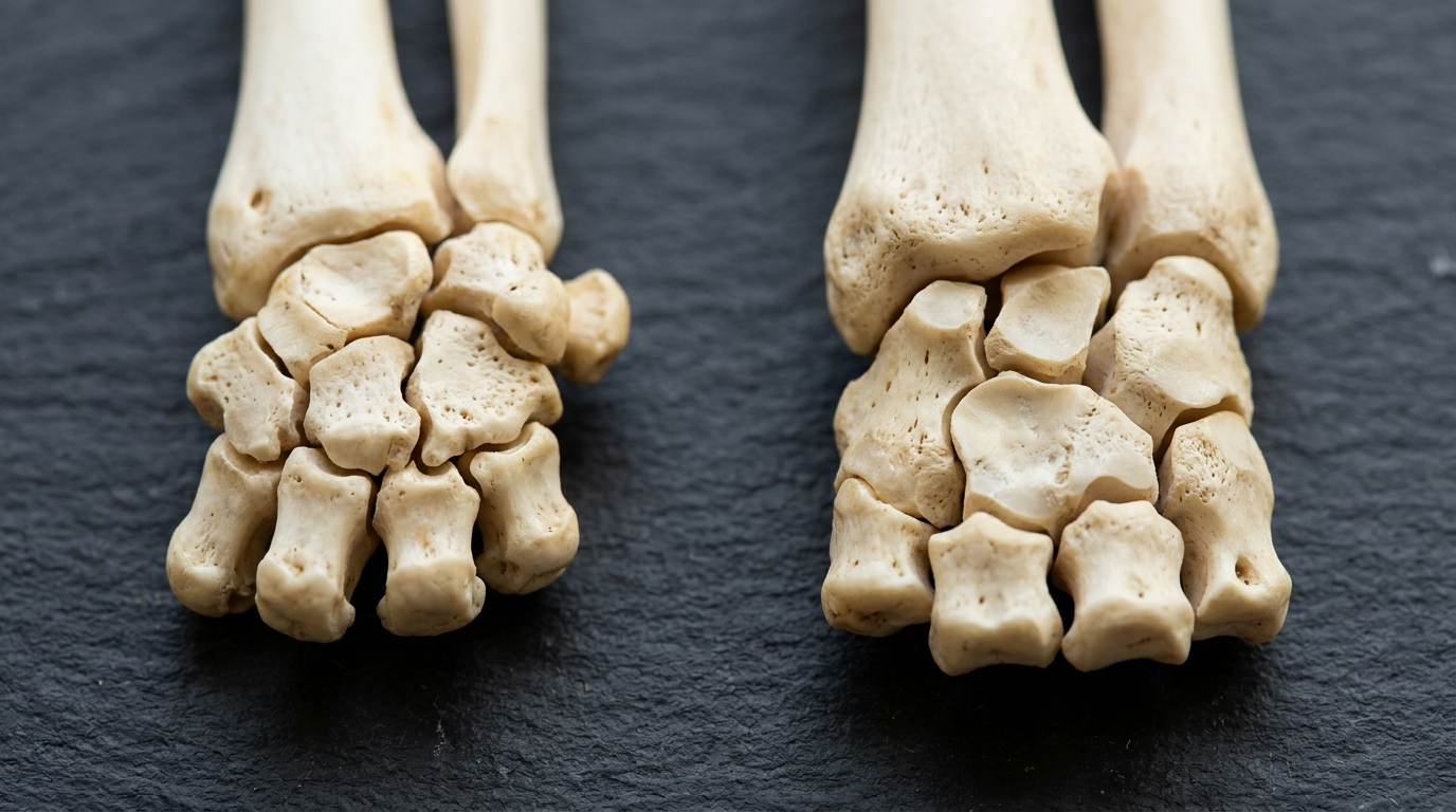
A comparative microcomputed tomographic investigation has revealed that domestic cats possess a markedly denser, thicker, and structurally distinct carpal bone architecture compared to toy breed dogs of matching body weight. In an examination of eighty carpal joints collected from twenty feline and twenty toy canine cadavers, researchers determined that feline carpal bones feature significantly higher bone volume fractions, thicker trabecular struts, and wider inter-trabecular spacing alongside considerably thicker cortical shells. Conversely, toy dogs exhibited a higher total count of individual trabeculae, substantially greater bone surface-to-volume ratios, and elevated connectivity densities across all seven carpal bones. Furthermore, no statistically significant structural variations were identified between the left and right limbs within either species.
Carpal joint injuries in cats present challenging clinical conditions characterized by low overall incidence rates between 0.26 and 0.29 percent, yet they carry substantial post-surgical complication frequencies ranging between 25 and 35 percent. Because carnivore carpal anatomy comprises seven distinct bones supported by complex ligamentous structures, severe traumatic injuries often involve simultaneous osseous fractures and ligament ruptures requiring temporary immobilization, ligament reconstruction, or surgical joint fusion through partial or total arthrodesis. Such surgical repairs typically necessitate the secure placement of metallic implants directly into one or more carpal elements. Prior research had extensively examined ligamentous discrepancies between canines and felines, but internal trabecular and cortical configurations across the complete carpal row had remained uncharacterized. Consequently, the research team designed this microcomputed tomographic evaluation to establish a reference baseline of feline and toy canine osseous microarchitecture, aiming to explain species-specific structural differences that could inform future veterinary orthopedic implant adaptations and fracture fixation strategies.
To eliminate body weight and size discrepancies as confounding factors, the study cohort comprised twenty cats and twenty toy breed dogs of approximately equal weight categories. All eighty evaluated joints were harvested from deceased or humanely euthanized animal subjects obtained without ante-mortem experimental intervention. The investigators specifically selected mature specimens with an estimated chronological age spanning eight to twelve years, an intentional demographic constraint aimed at minimizing age-related skeletal remodeling and microdamage while maintaining a balanced representation of neutered and sexually intact individuals. Because verified chronological birth records were unavailable, individual ages were systematically estimated through detailed dental status assessments and general external morphological examinations. The investigators enforced strict health criteria, excluding any candidate cadavers demonstrating pre-existing carpal fractures, joint degeneration, or radiographic evidence of osteoarthritis. Orthogonal two-plane radiographic screenings were conducted prior to freezing the specimens, employing a Siemens Luminos dRF diagnostic X-ray system configured at fifty kilovolts and 1.6 milliampere-seconds to verify joint integrity.
Following confirmation of structural joint normalcy, the cadaveric specimens were stored frozen at minus twenty-one degrees Celsius before undergoing a controlled two-day thawing period at ambient room temperature. Researchers carefully dissected away surrounding soft tissues while preserving the primary collateral and internal ligaments required to maintain correct anatomical alignment among the carpal bones. To ensure standardized sample positioning and container fitting, anatomical segments were cleanly transected two centimeters proximal to the joint within the antebrachium and two centimeters distal to the joint within the metacarpus, followed by fixation in four percent neutral formalin solution. High-resolution three-dimensional imaging was subsequently conducted on a Scanco Medical microcomputed tomography scanner, model micro-CT eighty, operating at tube settings of seventy kilovolts, one hundred fourteen microamperes, and an integration time of five hundred fifty milliseconds. Scans utilized a native isotropic voxel dimension of ten micrometers with one thousand angular projections captured over a one-hundred-eighty-degree rotation.
The three-dimensional scanning boundaries captured the entire carpal complex, comprising the intermedioradial, ulnar, accessory, and first through fourth carpal bones. Post-acquisition image processing was executed using proprietary microcomputed tomography evaluation software version 6.6. To analyze the cancellous compartment comprehensively, every tomographic slice sequence was inspected to identify the extreme proximal and distal boundaries containing trabecular structures, establishing a defined region of interest manually traced across each contiguous section. Because domestic cats and toy dogs exhibit distinct mineralization levels, overall bone dimensions, and grayscale intensity distributions, the research team instituted species-specific thresholding values to binarize grayscale datasets accurately into bone and non-bone voxels. Three independent experienced reviewers visually evaluated representative slices to establish threshold boundaries that delineated fine trabecular struts without including marrow spaces or producing partial volume artifacts. The arithmetic mean of these three independent evaluations was implemented as the final binarization threshold for each respective species, providing an empirical standard across all specimens.
Quantitative morphometric evaluation of the binarized trabecular volumes encompassed seven standardized metrics: bone volume fraction representing the percentage of segmented bone within the volume of interest; bone surface-to-volume ratio indicating architectural complexity; trabecular number derived as the reciprocal of mean distance between structural axes; trabecular thickness and trabecular separation quantified via three-dimensional sphere-fitting algorithms; connectivity density measuring trabecular junctions per unit volume; and degree of anisotropy assessing directional orientation. Cortical structure was classified by a single reviewer using a validated three-tier scheme on the middle third of each bone: score one designated interrupted bone lamellae, score two indicated continuous bone lamellae, and score three denoted normal, fully formed lamellar bone. The intermedioradial bone was further evaluated across its dorsal, palmar, and ulnar-facing surfaces, while the accessory bone was divided into articular and non-articular zones. General linear modeling was implemented in R software version 4.2.1, with normality, variance homogeneity, and heteroscedasticity evaluated prior to log transformations and Tukey-adjusted marginal mean comparisons.
The quantitative trabecular comparisons established highly significant morphological differences between the two companion animal species across all anatomical locations. Domestic cats demonstrated significantly higher bone volume fractions than toy dogs across all seven carpal bones, with statistical probabilities below 0.0097. In sharp contrast, toy dogs displayed significantly greater bone surface-to-volume ratios across every carpal element at probability values below 0.001, confirming a less compact trabecular network. Toy dogs also exhibited significantly higher trabecular numbers across all seven carpal bones compared to cats. Feline joints, however, displayed significantly greater trabecular thickness values across all bones, alongside significantly wider trabecular separation distances compared to their canine counterparts. Within the feline cohort, the ulnar carpal bone exhibited exceptional morphological density, presenting the highest bone volume fraction, the lowest surface-to-volume ratio, the thickest trabeculae, and significantly narrower trabecular spacing than either the intermedioradial or accessory carpal bones of the proximal carpal row.
Trabecular connectivity density was significantly elevated in toy dogs relative to cats across all seven carpal elements, with probability values below 0.001, while the accessory carpal bone demonstrated the lowest numerical connectivity values within both species groups. The degree of anisotropy was significantly greater in the feline accessory carpal bone compared to the canine equivalent, indicating a strongly aligned trabecular orientation aligned with directional forces, whereas differences across the remaining six carpal bones remained minor. Cortical bone evaluations revealed that cats consistently achieved maximum scores of three across all seven carpal bones, demonstrating evenly formed, continuous cortical shells. In contrast, toy dogs exhibited significantly diminished cortical development, frequently receiving scores of one characterized by discontinuous or interrupted bone lamellae, particularly across the distal carpal row and the dorsal surface of the intermedioradial bone. Both species maintained robust score-three cortical development on the palmar surface of the intermedioradial bone and at the muscular attachment sites of the accessory carpal bone.
The distinct morphological divergence between cats and toy dogs aligns with biomechanical principles of functional skeletal adaptation described by Wolff’s law, reflecting differences in natural physical behaviors and loading environments. Feline locomotion incorporates demanding mechanical activities including high-impact landing, climbing, and jumping, which exert substantial multi-axial stresses that stimulate cortical thickening and dense trabecular mineralization. Additionally, the feline carpus possesses an expansive range of pronation and supination measuring approximately one hundred fifteen degrees, compared to only fifty degrees in canines. Conversely, the attenuated cortices observed in toy dog breeds may stem from reduced mechanical loading, potentially exacerbated by owner handling behaviors such as frequent carrying. Furthermore, the specialized structure of the accessory carpal bone, functioning primarily as an apophysis under tensile loading rather than axial compression, accounts for its lower connectivity and highly anisotropic alignment.
Several methodological limitations were identified by the authors, primarily concerning the use of cadaveric specimens with estimated rather than precisely documented chronological ages. Although restricting the cohort to an estimated span of eight to twelve years minimized age-associated bone loss, and documented systemic illnesses like chronic renal insufficiency or hyperadrenocorticism were excluded based on available medical histories, undocumented subclinical factors cannot be entirely eliminated. Additionally, while utilizing species-specific image binarization thresholds accounted for inter-species differences in mineralization and voxel grayscale values, it introduced potential observer-dependent variability, though multi-reviewer averaging mitigated subjective bias. The researchers also noted that incorporating an intermediate group of medium-sized canines could provide additional comparative context in future investigations. Nonetheless, by confirming that all seven carpal bones possess cancellous architecture capable of supporting orthopedic implant fixation while demonstrating that feline bones possess superior bone volume fractions and thicker cortices, this investigation delivers critical microarchitectural benchmarks that can guide the engineering of species-tailored implants and improve surgical outcomes for carpal trauma.
Subject of Research: Microcomputed tomographic comparison of feline and canine carpal bone microarchitecture
Article Title: Microcomputed tomographic comparison of the bony structure of the canine and feline carpus
Article References: Goldstein, L., Waselau, A.-C., Zablotski, Y., & Meyer-Lindenberg, A. (2026). Microcomputed tomographic comparison of the bony structure of the canine and feline carpus. Acta Veterinaria Scandinavica, 68(1), Article 26. https://doi.org/10.1186/s13028-026-00871-8
Image Credits: AI Generated
DOI: 10.1186/s13028-026-00871-8
Keywords: veterinary orthopedics, microcomputed tomography, carpal joint, feline anatomy, canine anatomy, bone microarchitecture, trabecular bone, cortical bone, Microcomputed, tomographic, comparison, bony
News Source: William Thompson. (November 7, 2026). Microcomputed Tomography Reveals Significant Wrist Bone Differences in Cats and Dogs. Scienmag.