A systematic review published in BMC Artificial Intelligence synthesizes a decade of research on deep learning architectures applied to gigapixel whole-slide imaging in oncology, examining how computational pathology models evaluate digitized tissue specimens for cancer diagnosis and transcriptome profile prediction. Authored by Arfa Jabin, Jyoti Singh Kirar, and Shandar Ahmad, the comprehensive survey evaluated peer-reviewed studies published between January 2015 and May 2025 to trace the technological transformation of histological analysis from conventional microscopic inspection to automated predictive modeling. Histopathology historically relied on manual microscopic examination, chemical tissue preservation, and physical glass slide archiving, but the commercial emergence of whole-slide scanners in the late 1990s enabled complete slide digitization at extreme optical resolutions. Across the surveyed literature, the integration of artificial intelligence and computer vision contributed to a measurable diagnostic accuracy increase in digital pathology from 30 percent in 2013 to 53 percent in 2020, facilitating remote telepathology consultations and automated screening.
The digital pathology workflow begins with optical scanning platforms such as the Leica Aperio AT2, 3DHISTECH Pannoramic 250 Flash III, Hamamatsu NanoZoomer series, and the Philips IntelliSite Pathology Solution, which achieved primary diagnostic clearance from regulatory authorities. These scanners capture histological tissue sections at 20x or 40x magnification, producing gigapixel whole-slide image files that frequently reach dimensions such as 95,488 by 81,920 pixels. Because processing a gigapixel image directly through deep neural networks remains computationally infeasible, modern computational frameworks structure each whole slide into a multi-resolution image pyramid spanning several magnification levels. Level 0 represents 40x magnification for capturing intricate cellular morphology, Level 1 provides 10x magnification for tissue-level organization, Level 2 offers 2.5x magnification for broader structural patterns and region-of-interest identification, and Level 3 provides 1.25x magnification for rapid slide navigation. Open-source preprocessing pipelines like SliDL execute color normalization, artifact removal, and tissue segmentation, often utilizing Otsu thresholding to distinguish tissue foreground from glass background.
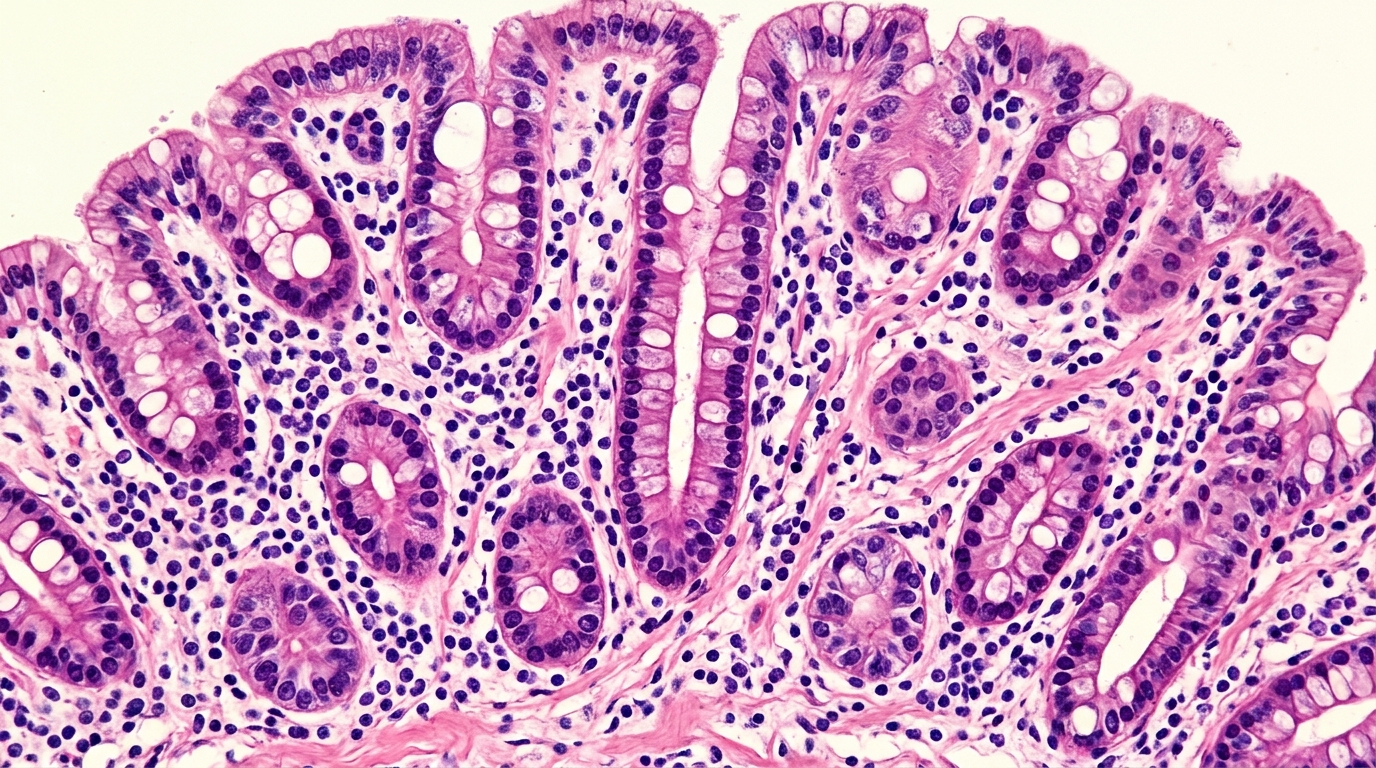
To translate cellular structures into machine-readable formats, early computational workflows applied quantitative image analysis to extract handcrafted mathematical measurements, including texture metrics derived from gray-level co-occurrence matrices, nuclear dimensions, and cell density. In comparative clinical benchmarks, quantitative image analysis models demonstrated tangible diagnostic value, such as a convolutional neural network achieving a positive predictive value of 0.71 and a negative predictive value of 0.96 when detecting invasive ductal carcinoma. Another quantitative model reached an area under the receiver operating characteristic curve of 0.97 in classifying prostate adenocarcinoma. These engineered approaches helped reduce subjectivity and inter-observer variability between pathologists, yet modern computational pathology has largely transitioned toward end-to-end deep learning. Contemporary deep learning architectures bypass manual feature engineering by learning predictive spatial patterns directly from raw image pixels. Pathologists distinguish normal tissue slides—which display preserved architecture, uniform staining, and clear white background spaces—from tumor slides that exhibit dense cell clusters, disrupted tissue morphology, and necrosis-related staining irregularities.
Because clinical datasets routinely provide only global slide-level diagnostic labels without painstaking patch-level boundary annotations, computational pathology relies heavily on weakly supervised multiple instance learning frameworks. In multiple instance learning, each whole slide functions as an aggregate bag composed of thousands of individual tile instances, typically sized at 256 by 256 or 512 by 512 pixels. Initial aggregation methods relied on simple mean or maximum pooling across tiles, but attention-based multiple instance learning introduced adaptive weighting mechanisms to spotlight diagnostically significant tissue regions while dampening uninformative background tiles. Subsequent innovations expanded this capability: clustering-constrained attention multiple instance learning separates high-attention from low-attention instances using only global slide labels, while Segment Anything Model-guided multiple instance learning incorporates zero-shot segmentation representations to sharpen spatial awareness. Dedicated interpretability models such as Attributable multiple instance learning explicitly assign slide-level diagnostic decisions back to individual tile features, generating visual attention heatmaps that pathologists can review.
Architectural evolution within multiple instance learning has moved beyond unordered patch sets to capture complex spatial context and tissue topology across whole slides. TransMIL treats image tiles as ordered sequences using transformer mechanisms to model long-range spatial dependencies, while the Whole-slide Image Transformer deploys a hierarchical architecture for local and global representation learning. Graph-based frameworks such as MulGT integrate graph neural networks with transformers to map topological connectivity between adjacent tissue patches, preserving localized spatial relationships. Meanwhile, dual-stream architectures like DSMIL and DTFD-MIL jointly optimize instance-level and bag-level representations to improve generalization across heterogeneous tissue morphologies, a paradigm further refined in DSMIL+ through self-supervised feature fusion. To balance extreme scale with computational efficiency, the Hierarchical Image Pyramid Transformer processes slides across tiered magnification levels, such as 20x, 10x, and 5x. Similarly, foundation frameworks like SAMPLER and contrastive pre-trained systems extract transferable representations from massive unlabeled pathology datasets with substantially lower computational expenditure.
Across 23 benchmarked studies examining 11 specific cancer types and two multi-cancer categories between 2008 and 2025, deep learning models demonstrated strong diagnostic classification performance, with convolutional neural networks and vision transformers reaching area under the curve metrics between 0.98 and 0.99. However, the survey revealed pronounced research concentration: breast cancer comprised 39 percent of analyzed literature across nine studies, and colorectal cancer represented 30 percent across seven studies. Models targeting breast cancer ranged from hybrid convolutional-recurrent architectures achieving 0.99 accuracy to multimodal transformer systems scoring 0.72 accuracy, while colorectal models achieved area under the curve values up to 0.99 through Phikon-v2 integration and self-supervised multiple instance learning. Gastric cancer and prostate cancer demonstrated consistent performance with area under the curve metrics of 0.98 and 0.92, respectively. Furthermore, pan-cancer foundation platforms like Virchow and CHIEF achieved generalizable area under the curve values between 0.91 and 0.94 across diverse tumor types.
To overcome the limitations of task-specific models trained on isolated institutional datasets, the field has increasingly adopted large-scale self-supervised foundation models that learn universal histological representations. A prominent example is GigaPath, a vision foundation model trained on 1.3 billion image tiles extracted from 171,189 whole-slide images representing more than 30,000 patients across 28 distinct cancer centers and 31 tissue types. Its derivative architecture, Prov-GigaPath, incorporates the LongNet architecture to perform whole-slide inference, achieving advanced performance in tumor detection, subtyping, immune cell quantification, and clinical prognosis estimation. Beyond standard brightfield hematoxylin and eosin slides, emerging diagnostic modalities are expanding the analytical boundaries of digital pathology. Multispectral and hyperspectral imaging capture granular biochemical spectral bands beyond visual light, reaching over 80 percent tissue differentiation accuracy during surgical procedures. Simultaneously, three-dimensional imaging modalities combining light-sheet microscopy and tissue clearing provide deep architectural volume analysis, overcoming traditional two-dimensional histology limitations through artificial intelligence-driven reconstruction.
A transformative development surveyed in the review is the computational prediction of transcriptome profiles directly from hematoxylin and eosin stained whole-slide images without sequencing tissue RNA. This predictive paradigm operates on the premise that underlying transcriptional activity and molecular properties govern observable histological morphology. The landmark HE2RNA framework used a pre-trained ResNet-50 network on 224 by 224 pixel tiles, aggregating 2048-dimensional feature vectors to predict bulk RNA sequencing profiles, achieving a Pearson correlation coefficient of 0.79 for inflammatory genes and an area under the curve of 0.92 for microsatellite instability. Subsequent architectures introduced specialized improvements: SEQUOIA utilized a linearized transformer across 7,584 tumor samples from 16 cancer types to predict 7,851 genes; tRNAsformer combined multiple instance learning with transformers, achieving a mean squared error of 0.18 and a Pearson correlation of 0.84; and PRALINE incorporated prior biological gene-gene interaction networks, increasing well-predicted genes by 983 in breast cancer.
Additional transcriptomic frameworks have expanded molecular mapping capabilities, including hist2RNA for patient-level breast cancer subtype identification, BrST-Net for resolving spatial tumor heterogeneity, and DL-MIL for predicting the 21-gene OncotypeDx recurrence panel with an area under the receiver operating characteristic curve of 0.831. At the pan-cancer scale, a deep regression model trained on approximately 10,000 tumors across 20,530 genes achieved a Pearson correlation of 0.83 for its top 1,000 genes with an 0.18 mean squared error. In spatial transcriptomics, the Spatial Transcriptomics Graph Attention Network estimates spot-level expression, SpaCKLE imputes missing expression measurements, and contrastive HECLIP aligns histological representations with transcriptomic profiles. Furthermore, multimodal large language models such as GPT-4 and GPT-4V demonstrate diagnostic capabilities, with GPT-4 achieving 84 percent accuracy in neuropathology assessments and multimodal systems matching specialized medical imaging models when synthesizing visual histology with textual clinical reporting.
Despite impressive benchmark metrics, the review identifies critical barriers that must be resolved prior to routine clinical translation. Primary among these is the validation gap: many algorithms suffer from staining variability, scanner batch effects, and lack of external validation across multi-institutional cohorts, leading to potential overfitting on academic populations. In transcriptomic prediction, biological validation remains incomplete, raising concerns over whether models learn genuine regulatory mechanisms or capture statistical artifacts. Furthermore, models like SEQUOIA demand substantial computing resources, requiring 80 gigabytes or more of GPU memory, which limits deployment in resource-constrained clinics. Future progress relies on privacy-preserving federated learning across hospital networks, multimodal integration combining whole-slide imaging with radiology and clinical records, and rigorous prospective clinical trials directly comparing algorithm-guided outcomes against standard diagnostic assays. Addressing these challenges through standardized imaging protocols and explainable artificial intelligence will determine whether computational pathology successfully transitions into an actionable predictive discipline.
Subject of Research: Deep learning models for cancer diagnosis and transcriptome prediction from whole-slide imaging
Article Title: AI-based methods for modelling whole-slide imaging data in cancer diagnosis and transcriptome profile prediction
Article References: Jabin, A., Kirar, J. S., & Ahmad, S. (2025). AI-based methods for modelling whole-slide imaging data in cancer diagnosis and transcriptome profile prediction. BMC Artificial Intelligence, 1(1), Article 16. https://doi.org/10.1186/s44398-025-00018-1
Image Credits: AI Generated
DOI: 10.1186/s44398-025-00018-1
Keywords: digital pathology, whole slide imaging, deep learning, cancer diagnosis, transcriptome prediction, multiple instance learning, vision transformers, computational pathology, AI-based, methods, modelling, whole-slide
News Source: Juliet Wilcox. (November 6, 2026). Artificial Intelligence Models Predict Cancer Diagnoses and Gene Expression from Tissue Slides. Scienmag.