A fully automated, artificial intelligence-powered biophysical model has outperformed the standard manufacturer-provided estimates at predicting the volume of tissue destroyed during radiofrequency ablation of liver cancer, according to a retrospective clinical evaluation published in European Radiology Experimental. In a cohort of 96 ablation procedures performed in 65 patients with hepatocellular carcinoma, the patient-specific simulation achieved a median relative volume difference of 0.39 from the measured ablation zone, compared with 1.04 for the manufacturer’s approximation, a statistically significant improvement.
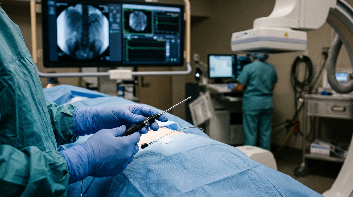
Radiofrequency ablation is an established, minimally invasive treatment for hepatic malignancies. The procedure involves image-guided percutaneous needle insertion into the tumor to achieve heat-induced tissue necrosis. Successful ablation requires tumor safety margins of at least 5 mm to avoid tumor residue or recurrence, which depends heavily on the operator’s experience in choosing the type and number of needles, their placement, and the appropriate ablation settings such as power and duration.
Predicting the final ablation volume to ensure sufficient margins remains challenging. Needle manufacturers provide expected ablation zones for specific settings, but these are typically spherical or ellipsoidal approximations derived from idealized ex vivo data. Several patient-specific factors, including the condition of the liver parenchyma, blood perfusion, and proximity to large vessels, significantly influence the outcome. The heat-sink effect, particularly relevant when large vessels with a diameter greater than 5 mm lie close to the ablation zone, can reduce efficacy and cause local recurrence, because flowing blood carries heat away from the target tissue.
The new study, conducted at a Swiss center on a prospectively collected patient cohort from 2011 to 2022, evaluated a model that simulates the physical mechanisms underlying the procedure using each patient’s own anatomy and procedural data. The retrospective analysis included 96 radiofrequency ablation procedures in 65 patients treated with a single ablation using a cluster needle. Tumors, needle position, and the post-ablation zone were manually segmented on periprocedural computed tomography scans, while the liver parenchyma and vascular structures were extracted automatically using deep learning.
The biophysical model, accelerated on a graphics processing unit, simulated heat transfer through hepatic tissue using the bioheat equation, incorporating the cooling effect of major blood vessels through a vessel indicator function that applies the reaction term only within segmented hepatic blood vessels. A three-state necrosis model computed the concentration of alive, vulnerable, and dead cells over time based on the simulated temperature, assuming that cells stop functioning if the temperature exceeds a threshold for a sufficient duration and recover if the threshold is not maintained long enough. The heat source was modeled with an ellipsoidal boundary condition centered at the needle tip, calibrated so that in a vessel-free ex vivo scenario the predicted zone matched the manufacturer’s specification of an ellipsoid with a long axis of 45 mm and a short axis of 42 mm.
The clinical evaluation first tested whether simple, manually measured parameters could predict the ablation volume. The researchers examined the distance between the tumor and the liver capsule, the distance to the closest vessel, the type and diameter of that vessel, the final needle-tip temperature, and the ablation time. None of these parameters showed a statistically significant correlation with the final ablation volume, with Pearson correlation coefficients ranging from -0.022 to 0.29. Even under a standardized ablation protocol in a similar patient cohort, the final ablation volumes varied widely, from 7.9 to 78.8 cubic centimeters, with a median of 24.07 cubic centimeters.
The model’s predictions, by contrast, tracked the actual outcomes closely. Across 94 evaluable cases, the simulated and ground-truth ablation zone volumes were statistically similar, while the manufacturer estimates significantly overestimated the ablation volume. The model achieved a median relative volume difference of 0.39, with an interquartile range of 0.24 to 0.57, significantly lower than the manufacturer’s algorithm at 1.04, with an interquartile range of 0.64 to 1.61. A relative volume difference of 0.30 corresponds to a 30 percent volume difference, so the manufacturer’s figure represents errors exceeding 100 percent in the typical case. Volume similarity analysis showed no systematic over-ablation or under-ablation by the model, except in cases where the ablation time was unknown, when both the model and the manufacturer overestimated the post-RFA ablation volume.
Incorporating patient-specific ablation time further improved accuracy. Ablation durations were available for 87 of the 96 procedures, with a median ablation time of 420 seconds, or 7 minutes, ranging from about 3 minutes to 42 minutes. For the nine procedures with unknown ablation time, the researchers assumed the manufacturer’s standard 12-minute duration. The center does not enforce the 12-minute limit, instead waiting for three roll-offs, points at which the impedance-controlled generator automatically shuts off when it detects a significant increase in impedance. The total ablation time is documented in the ablation report, but the precise duration and timing of each roll-off is not reported and therefore could not be considered in the analysis.
Speed is a notable feature of the system. The mean computational time to simulate 1 second of ablation was 0.45 seconds, with a 95 percent confidence interval of 0.42 to 0.47, on an NVIDIA RTX A4500 GPU, using a Lattice Boltzmann Method to solve the heat transfer equations. The pipeline is fully automated because it relies on a deep learning segmentation method, eliminating manual steps prone to human error and allowing direct analysis of intra-procedure data rather than diagnostic images, without complex image registration. Manual processing of a single ablation case for validation purposes required approximately 30 minutes, encompassing annotation of the needle position and segmentation of both the tumor and the ablation zone.
The authors note that this is, to their knowledge, the first report focused on hepatocellular carcinoma with a patient cohort reflecting real-world clinical conditions. The included patients had underlying liver fibrosis or cirrhosis, producing heterogeneous liver parenchyma that differs significantly from the healthy, homogeneous, non-perfused liver tissue typically used in ex vivo studies by device manufacturers. While the current model still relies on nominal parenchymal tissue properties rather than fibrosis- or cirrhosis-specific values, it nonetheless yielded substantially more accurate volume predictions than the manufacturer reference. The researchers expect that explicitly incorporating fibrosis- and cirrhosis-dependent tissue properties will further improve prediction accuracy, a focus of future work. Two cases were excluded from the model evaluation due to poor liver segmentation quality, one involving a major right hepatectomy and the other a lipiodol injection, scenarios absent from the deep learning algorithm’s training data.
The study has limitations the authors themselves identify. It focused on predicting ablation volume rather than spatial extent, because evaluating the spatial distribution would require elastic registration between pre-procedure and post-procedure CT images, and no robust, fully automatic registration algorithms tailored to ablation data are currently available. The use of nominal tissue parameters also does not reflect inter-patient variability in hepatic perfusion or tissue heterogeneity. The authors state that a key next step will be to evaluate whether model performance correlates with clinically meaningful outcomes such as local tumor progression, recurrence-free survival, and ablative margin adequacy, and that the intended next step is to deploy the model in a preoperative planning setting using pre-procedural imaging, where it could recommend an optimal ablation strategy including the number of needles and their positioning, with the goal of achieving complete tumor coverage while minimizing damage to surrounding healthy tissue.
Subject of Research: An AI-powered patient-specific biophysical model for predicting radiofrequency ablation volumes in hepatocellular carcinoma
Article Title: Patient-specific biophysical AI-powered modeling for predicting RFA volumes in HCC
Article References: Audigier, C., Akkari, F. G., Meister, F., Tonglet, A., Maréchal, B., Frings, O., & Duran, R. (2026). Patient-specific biophysical AI-powered modeling for predicting RFA volumes in HCC. European Radiology Experimental, 10(1), Article 166. https://doi.org/10.1186/s41747-026-00839-3
Image Credits: AI Generated
DOI: 10.1186/s41747-026-00839-3
Keywords: radiofrequency ablation, hepatocellular carcinoma, biophysical modeling, artificial intelligence, deep learning, heat-sink effect, liver cirrhosis, treatment planning, computed tomography, ablation volume prediction, GPU simulation, interventional radiology
News Source: Nathaniel Bowman. (November 6, 2026). AI model predicts liver tumor ablation volumes better than manufacturer estimates. Scienmag.