A 70-year-old man with an underlying interstitial lung disease arrived at the hospital with a week of cough, purulent sputum, fever, and right-sided pleuritic chest pain. High-resolution computed tomography revealed the cause: a fat-density lesion measuring approximately −100 Hounsfield units lodged inside his right middle lobe bronchus, completely blocking the airway and producing post-obstructive atelectasis and consolidation, the pattern known as middle lobe syndrome. Bronchoscopic biopsy confirmed the rarest of culprits, an endobronchial lipoma, a benign tumor of fat tissue that accounts for roughly 0.1% to 0.5% of pulmonary neoplasms. Even more unusual was its location, since fewer than ten cases of right middle lobe involvement have been reported in the English literature.
The case, published in Respirology Case Reports, carries a technical lesson that goes beyond its rarity. The patient’s medical history included combined pulmonary fibrosis and emphysema, a form of interstitial lung disease in which fibrotic changes in the lower lobes coexist with emphysema in the upper lobes. His high-resolution CT showed basal-predominant reticulation, traction bronchiectasis, and honeycombing consistent with a definite usual interstitial pneumonia pattern, diagnosed radiologically without lung biopsy. Because surgical resection in such patients carries high risk due to limited respiratory reserve, the treating team chose a lung-sparing bronchoscopic approach, removing the tumor with an electrocautery snare and ablating its base with argon plasma coagulation, avoiding surgery entirely.
What distinguishes this report is the documentation of pulmonary function before and after intervention, something the authors say has not previously been described for endobronchial lipoma treatment in patients with interstitial lung disease. Earlier historical testing from March and April 2022 had recorded FVC of 3.00 L, or 97% predicted, FEV1 of 2.32 L, or 102% predicted, an FEV1/FVC ratio of 77.3%, and a hemoglobin-corrected DLCO of 12.16 mL/min/mmHg, or 58% predicted. A later pre-event reference assessment, performed roughly three to six months before bronchoscopy when the patient was already clinically unwell with recurrent respiratory infections, showed FVC of 86% predicted and DLCO of 49% predicted.
During the year before admission, the man suffered recurrent respiratory infections localized to the right middle lung field. In the final six months, he experienced two exacerbations attributable to post-obstructive infection, each requiring antibiotics, systemic corticosteroids, and supplemental oxygen, with one requiring hospitalization. Repeat testing over this period documented a decline to FVC of 75% predicted and DLCO of 35% predicted. On examination, clinicians heard crackles and rhonchi over the right middle lung field alongside bilateral basal Velcro-like crackles typical of his fibrotic disease. Notably, the endobronchial obstruction had not been recognized on imaging during the previous hospitalization, when the appearances were attributed to post-obstructive consolidation alone, a recognized diagnostic pitfall that contributed to a delay in diagnosis.
Diagnostic flexible bronchoscopy revealed a smooth, yellowish-brown, polypoid, soft, and avascular lesion causing complete luminal occlusion at the carina between the RB4 and RB5 segmental bronchi. Initial forceps biopsy showed mature adipocytes covered by a normal columnar respiratory epithelial lining, with no cytological atypia and no lipoblasts, findings that excluded well-differentiated liposarcoma.
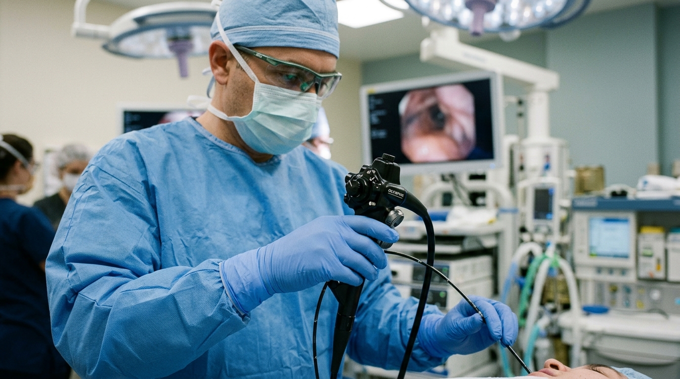
Therapeutic bronchoscopy was performed the next day as a flexible procedure under general anesthesia via a laryngeal mask airway. The team selected flexible over rigid bronchoscopy because the lesion sat distally at the RB4/RB5 carina and was soft and avascular on initial inspection, making it amenable to flexible instrumentation, while the laryngeal mask preserved ventilation and instrument access. The bulk of the lesion was resected with an electrocautery snare using EndoCut mode at 40 watts, effect 2, and a cut duration of 2, and the residual broad base was ablated with argon plasma coagulation at 40 watts with a 1 L/min flow. Mild, self-limiting hemorrhage at the base after ablation was controlled with suction alone. After resection, copious purulent secretions were aspirated from the airway distal to the obstruction and sent for microbiological analysis, which grew Pseudomonas species and anaerobes, a pattern the authors interpret as characteristic of chronic infection trapped behind an obstructing lesion rather than typical community-acquired pneumonia.
Complete airway patency was restored. At the two-month follow-up, a interval chosen to allow resolution of post-obstructive inflammation and consolidation while remaining within the patient’s established interstitial lung disease surveillance schedule, the patient was asymptomatic with radiological resolution of the obstruction confirmed on CT. Pulmonary function had climbed toward the later pre-event reference values: FVC rose from 75% to 83% predicted, an absolute gain of 8 percentage points, and DLCO rose from 35% to 50% predicted, a gain of 15 percentage points. The authors attribute this improvement to recanalization, drainage of retained secretions, and concurrent treatment of infection, while cautioning that the temporal pattern supports a potentially reversible component associated with the post-obstructive episode but does not establish that bronchoscopic intervention alone caused the improvement or exclude fluctuation or progression of the underlying interstitial lung disease.
The discussion in the report is careful about causality. Bronchoscopic resection directly reopened the mechanically occluded bronchus and allowed drainage of retained purulent secretions, which the authors regard as the central anatomical effect of treatment and a plausible important contributor to recovery. Yet suction and drainage, antibiotic and supportive treatment, resolution of infection and atelectasis, and ordinary measurement variability, particularly for DLCO, may all have contributed, and their individual effects cannot be quantified in a single case. The interval decline between the 2022 historical assessments and the later pre-event reference, from FVC 97% to 86% predicted and DLCO 58% to 49% predicted, may partly reflect the evolving obstruction and recurrent infection, but progression of the underlying interstitial lung disease over that interval cannot be excluded from the available measurements. The report also does not establish that the surveillance frequency of pulmonary function testing every two to three months shortened the diagnostic delay.
Several limitations frame the findings. The follow-up period was only two months, and although recurrence rates after bronchoscopic resection of endobronchial lipomas are not well characterized, incomplete base ablation could predispose to late recurrence occurring months to years after intervention, so longer surveillance with serial bronchoscopy and imaging is required to confirm durability. Completeness of base ablation was assessed visually rather than histologically verified. As a single case, generalizability is uncertain across the interstitial lung disease subtype, since combined pulmonary fibrosis and emphysema may respond differently to post-obstructive relief than other forms such as nonspecific interstitial pneumonia or hypersensitivity pneumonitis; across lesion characteristics, since fibrous or vascular lesions may require alternative techniques; and across patient factors, since the patient’s preserved FVC of 75% predicted may have facilitated recovery in ways that more severe restriction would not.
The clinical takeaway is straightforward. Endobronchial lipomas typically arise in large bronchi where adipose tissue is present, and although benign, they can cause progressive airway obstruction and distal lung damage, including bronchiectasis and irreversible parenchymal injury, when diagnosis is delayed. CT imaging plays an important role when fat attenuation is identified within an endobronchial lesion, but interpretation may be difficult in the presence of post-obstructive consolidation, and bronchoscopy remains the gold standard for direct visualization, histological confirmation, and therapeutic intervention. The authors suggest that maintaining a low threshold for bronchoscopy in persistent or recurrent lobar disease could prevent such delays, and they hypothesize that earlier intervention might further limit distal damage in cases where obstruction persists longer or infection is more severe, a proposition they note warrants prospective evaluation. For this patient, the lung-sparing procedure achieved airway reopening without surgical resection, a meaningful outcome in someone whose fibrotic and emphysematous lungs left little reserve to spare, though longer follow-up and additional cases are needed to assess how durable and generalizable the approach proves to be.
Subject of Research: Bronchoscopic removal of a rare right middle lobe endobronchial lipoma causing middle lobe syndrome in a patient with interstitial lung disease
Article Title: A Rare Endobronchial Lipoma of the Right Middle Lobe Causing Middle Lobe Syndrome and Recurrent Post‐Obstructive Infection in a Patient With Interstitial Lung Disease, Managed by Lung‐Sparing Bronchoscopy
Article References: Palamidas, A., Papatheodosiou, K., Karagianni, E., Daglis, F., & Bouros, D. (2026). A Rare Endobronchial Lipoma of the Right Middle Lobe Causing Middle Lobe Syndrome and Recurrent Post‐Obstructive Infection in a Patient With Interstitial Lung Disease, Managed by Lung‐Sparing Bronchoscopy. Respirology Case Reports, 14(11), Article e70800. https://doi.org/10.1002/rcr2.70800
Image Credits: AI Generated
DOI: 10.1002/rcr2.70800
Keywords: endobronchial lipoma, middle lobe syndrome, interstitial lung disease, bronchoscopy, combined pulmonary fibrosis and emphysema, pulmonary function testing, argon plasma coagulation, post-obstructive infection, airway obstruction, case report, Rare, Endobronchial
News Source: Ophelia Keating. (November 6, 2026). Rare Airway Tumor Choking Lung Lobe Removed by Bronchoscopy. Scienmag.