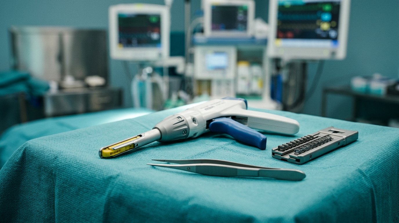
A clinical report published in General Thoracic and Cardiovascular Surgery Cases documents an extraordinary, life-threatening case of hemorrhagic shock resulting from eighth intercostal artery perforation caused by a staple leg protruding through reinforcement material after thoracoscopic pulmonary wedge resection in a 76-year-old male patient. Pulmonary wedge resection is an established minimally invasive lung-sparing surgical procedure widely utilized for both the diagnosis and treatment of pulmonary lesions, with mechanical staplers and staple line reinforcement systems playing a central role in optimizing surgical feasibility and suppressing postoperative air leakage. While staple line reinforcement sheets are routinely integrated into thoracic procedures to enhance mechanical sealing, their protective capacity against sharp mechanical vascular trauma has remained largely undefined in clinical literature. Vascular injuries stemming directly from surgical staple lines represent exceptionally rare but catastrophic complications within thoracic surgery, typically occurring when rigid metallic components interact abrasively with nearby cardiovascular structures. In this newly described surgical case, an exposed staple leg penetrated through the synthetic reinforcement backing and punctured the eighth intercostal artery, triggering severe hypovolemic shock that was dynamically precipitated by abrupt postoperative lung collapse and subsequent rapid re-expansion.
The patient presented with an extensive oncological history, having initially received a diagnosis of right upper lobe lung adenocarcinoma approximately five years and six months prior to the acute surgical event. That primary tumor had been classified as stage IVB disease (clinical T4N3M1c according to the eighth edition of the TNM classification) characterized by extensive metastatic involvement, including confirmed pleural dissemination, bilateral adrenal metastases, intra-abdominal lymph node metastases, and splenic metastasis. Molecular companion diagnostic profiling performed at the time of initial diagnosis revealed the presence of a KRAS G12C mutation alongside a programmed death-ligand 1 (PD-L1) tumor proportion score of 100 percent. Systemic treatment commenced with three cycles of combination chemoimmunotherapy consisting of cisplatin, pemetrexed, and pembrolizumab; however, this initial regimen was discontinued when the patient developed Common Terminology Criteria for Adverse Events grade 3 diarrhea. Following the resolution of gastrointestinal toxicity, the patient resumed therapy with eight cycles of pembrolizumab monotherapy, which subsequently had to be discontinued after the emergence of grade 2 immune-related pneumonitis and cutaneous rash. Corticosteroid management with oral prednisolone was initiated at 20 milligrams per day and gradually tapered over several months, successfully achieving and maintaining a complete clinical response for four consecutive years without additional cancer therapy.
Routine surveillance computed tomography conducted four years following the cessation of immune checkpoint inhibitor therapy revealed a new, distinct solid subpleural nodule measuring 14 by 10 millimeters situated within segment 6 of the right lower lobe. Preoperative axial and sagittal computed tomography scans demonstrated that the solitary lesion was located immediately adjacent to the visceral pleural surface. Complementary positron emission tomography integrated with computed tomography identified prominent metabolic activity within the solitary nodule, yielding a maximum standardized uptake value of 4.7. These diagnostic imaging characteristics raised immediate clinical concern for either an isolated oligometastasis or a metachronous primary bronchogenic carcinoma. Because the patient had achieved remarkable long-term disease-free survival from stage IVB disease and carried a documented vulnerability to immune-related pneumonitis, the multidisciplinary thoracic team prioritized a tissue-sparing, minimally invasive diagnostic approach. Thoracoscopic pulmonary wedge resection was accordingly planned to achieve definitive histologic identification and complete local excision of the segment 6 lesion while deliberately conserving functional surrounding lung parenchyma.
The thoracic operation was conducted under general anesthesia utilizing single-lung ventilation with the patient positioned in the left lateral decubitus orientation. Surgical access was established by placing a 12-millimeter port in the seventh intercostal space along the anterior axillary line, allowing direct visual identification of the target nodule in segment 6, followed by a utility incision placed in the sixth intercostal space along the posterior axillary line. Visual and tactile inspection revealed thin, compliant pulmonary parenchyma surrounding the peripheral lesion. To minimize post-resection air leakage, the surgical team performed the wedge resection using an ECHELON ENDOPATH Staple Line Reinforcement system utilizing a 60-millimeter gold cartridge, with cartridge height chosen based on manual assessment of tissue characteristics while accounting for the additional thickness of the reinforcement strip. Following tissue division, immediate inspection of the staple line showed satisfactory staple formation without visual malformations, and underwater submersion sealing tests demonstrated no evidence of air leakage or protruding staple legs. After resuming bilateral lung ventilation, the surgical team re-evaluated the staple line, verified integrity, placed a chest tube, and concluded the procedure. Subsequent pathological analysis confirmed a metachronous primary lung adenocarcinoma distinct from his previous malignancy, without evidence of interstitial lung disease.
The early postoperative course took an unexpected turn on postoperative day 1 when the patient experienced an episode of severe agitation and inadvertently removed his thoracic chest drain. Surgical staff immediately closed the chest tube insertion site, and an emergent chest radiograph identified mild subcutaneous emphysema without radiographic evidence of parenchymal lung collapse. By postoperative day 2, however, serial chest radiography revealed notable progression of subcutaneous emphysema across the chest wall, signaling persistent alveolar air leakage from the operative site. To manage the ongoing air leak and restore intrapleural pressure, clinicians promptly reinserted a thoracic chest tube, which successfully evacuated the intrapleural space and achieved complete parenchymal re-expansion. While the patient initially appeared stable following drain replacement, the dramatic sequence of mechanical lung deflation followed by rapid reinflation induced significant positional and spatial alterations within the thoracic cavity, setting the stage for subsequent vascular trauma.
On postoperative day 3, the patient experienced a sudden, catastrophic clinical deterioration marked by an abrupt loss of consciousness and profound cardiovascular collapse. Vital signs showed marked tachycardia with a heart rate of 110 beats per minute and profound hypotension with systolic blood pressure plummeting to 80 millimeters of mercury. Concurrently, the thoracic drainage system rapidly evacuated 500 milliliters of fresh blood, signaling acute and severe intrathoracic hemorrhage. Although an immediate laboratory assessment recorded a hemoglobin concentration of 10.5 grams per deciliter, the surgical team correctly recognized that circulating hemoglobin values frequently remain deceptively normal during the hyperacute phase of massive vascular hemorrhage before hemodilution manifests. Recognizing hypovolemic shock, clinicians transferred the patient for emergency re-exploration. During anesthesia induction, the patient suffered pulseless electrical activity cardiac arrest secondary to profound intravascular volume depletion. Advanced cardiopulmonary resuscitation was instantly initiated, including intravenous adrenaline administration and rapid blood component transfusions, successfully restoring spontaneous circulation within four minutes and permitting emergency thoracotomy.
Upon performing an emergency thoracotomy, the surgical team observed substantial hematoma formation clustered primarily throughout the posterior basal pleural cavity. Following rapid evacuation of the pooled blood and clots, active pulsatile arterial hemorrhage was directly visualized emerging from the eighth intercostal artery, which was temporarily controlled utilizing electrocautery. Thorough inspection of the hemithorax revealed an absence of pre-existing pleural adhesions, while direct examination of the bleeding intercostal artery demonstrated that its vascular walls possessed normal structural integrity without underlying inflammation, atherosclerotic fragility, or macroscopic pathology. Detailed examination of the nearby pulmonary resection margin identified a single metallic staple leg protruding through the synthetic staple line reinforcement material. Under controlled lung inflation, the operative team definitively observed that this exposed staple leg came into direct mechanical alignment and forceful contact with the bleeding eighth intercostal artery, proving that the protruding metal had perforated the vessel wall during pulmonary re-expansion.
To eliminate the source of mechanical irritation and achieve definitive surgical repair, the protruding staple leg was meticulously excised from the resection margin, and the resulting visceral pleural defect was closed using manual suture repair. Although subsequent sealing tests demonstrated complete absence of air leakage from the repaired lung parenchymal margin, the operating team applied an additional polyglycolic acid sheet (Neoveil) and fibrin glue over the entire staple line to provide robust coverage against future mechanical friction. Complete and secure vascular hemostasis was accomplished by ligating the eighth intercostal artery both proximally and distally to the perforation site, permanently arresting hemorrhage. Total intraoperative blood loss, including the evacuated intrapleural hematomas, measured 940 milliliters. Following this complex revision surgery, the patient experienced an uneventful recovery with no further bleeding or respiratory complications, culminating in successful hospital discharge on postoperative day 10.
In evaluating the broader clinical implications of this incident, the reporting authors stressed that direct mechanical vascular injury provoked by staple protrusion represents an exceedingly rare but critical complication in thoracic surgery. Although previous medical literature has documented staple-related complications including intercostal artery laceration, late-onset hemothorax, and aortic wall erosion, the vast majority of historical reports involved standard, unreinforced staple lines where prolonged, low-grade mechanical friction caused delayed vascular perforation weeks or months later. This case is uniquely instructive because it represents one of the few recorded instances of intercostal artery perforation occurring despite the proactive deployment of a reinforced stapler system, demonstrating that synthetic reinforcement sheets do not provide an impenetrable barrier against rigid metallic staple malformation. The authors determined that the sudden shift in anatomical geometry—sparked by lung collapse after chest tube avulsion followed by forced re-expansion—drove the protruding staple directly into the adjacent chest wall, a vulnerability heightened by the greater mobility and displacement range of the lower pulmonary lobe.
The case report further highlights essential technical factors governing surgical cartridge selection and the physical mechanics of tissue compression in reinforced stapling. In this patient, a gold cartridge was selected based on visual and tactile evaluation of the thin pulmonary parenchyma; however, adding reinforcement material increases effective compressed tissue thickness, which can impede complete B-shaped staple formation and prevent adequate leg closure. Because subtle staple malformations may escape detection during routine visual inspection under resting intraoperative conditions, subsequent respiratory cycling, pulmonary compliance shifts, and rapid reinflation can distort staple line geometry and expose sharp metallic edges. The authors emphasized that staple formation depends on complex interactions among tissue thickness, compressibility, and cartridge staple height rather than subjective thickness assessment alone. Consequently, surgical teams must conduct exhaustive intraoperative inspections of staple lines, consider trimming or covering suspicious metallic edges—especially near the chest wall or on mobile lobes—and maintain rigorous chest drain protocols to avoid abrupt lung volume changes that could precipitate catastrophic mechanical injuries.
Subject of Research: Mechanical intercostal artery injury from staple protrusion following reinforced pulmonary wedge resection
Article Title: Life-threatening hemorrhagic shock due to intercostal artery perforation from a protruding staple leg after pulmonary wedge resection with a reinforced stapler: a case report
Article References: Watanabe, S.-N., Kimura, D., Sasaki, T., Kimura, S., Ichinohe, C., Kato, T., Ono, S., Imai, H., & Minakawa, M. (2026). Life-threatening hemorrhagic shock due to intercostal artery perforation from a protruding staple leg after pulmonary wedge resection with a reinforced stapler: a case report. General Thoracic and Cardiovascular Surgery Cases, 5(1), Article 24. https://doi.org/10.1186/s44215-026-00256-3
Image Credits: AI Generated
DOI: 10.1186/s44215-026-00256-3
Keywords: pulmonary wedge resection, surgical stapler, staple line reinforcement, intercostal artery perforation, hemorrhagic shock, thoracoscopic surgery, case report, postoperative complications, Life-threatening, hemorrhagic, shock, intercostal
News Source: Ophelia Keating. (November 6, 2026). Protruding Staple Leg Causes Severe Intercostal Artery Bleeding After Lung Resection. Scienmag.