Cardioembolic stroke, in which a thrombus or other material originating in the heart travels to the brain, accounts for roughly 15 to 40 percent of all ischemic strokes, and a new systematic review argues that finding its source demands more than an imaging checklist. The review, published in Cardiovascular Ultrasound, maps the full spectrum of cardiac and aortic substrates that can seed cerebral embolism and lays out a scenario-based diagnostic strategy built around echocardiography, with cardiac computed tomography and cardiac magnetic resonance reserved for selected cases. Its central message is that a clinically driven, patient-specific work-up, rather than indiscriminate extensive testing, is what improves diagnostic yield while avoiding unnecessary investigations.
The stakes are considerable. Stroke remains one of the leading causes of death and long-term disability worldwide, and although age-adjusted mortality has declined in high-income regions, the absolute number of cases continues to rise with population ageing and growth. In 2021, approximately 94 million people were living after a stroke and nearly 12 million new stroke events occurred globally, with projections indicating further substantial increases by 2050, particularly in low- and middle-income countries where prevention programs and access to care remain limited. Ischemic stroke makes up about 87 percent of all strokes, and cardioembolic strokes tend to produce larger infarcts, more profound neurological deficits, and a higher early recurrence risk than non-cardioembolic subtypes, which makes early identification of the cardiac source crucial.
The range of potential substrates is wide. Atrial fibrillation is the dominant one, but native and prosthetic valvular disease, left ventricular thrombus, aortic arch atheroma, intracardiac tumors, and right-to-left shunts such as patent foramen ovale all contribute significantly to risk. The review describes how clinicians can suspect a cardioembolic mechanism from clinical clues: maximal deficit at onset, cortical symptoms such as aphasia or neglect, evidence of systemic embolism, and a history of atrial fibrillation, recent myocardial infarction, heart failure, valvular disease, or cardiac masses. Neuroimaging strengthens the case when it shows cortical or cortico-subcortical infarcts, multiple acute lesions in different vascular territories, wedge-shaped territorial infarcts, or large subcortical lesions inconsistent with lacunar disease, and vascular imaging may reveal abrupt occlusion without significant stenosis. No single feature is diagnostic in isolation, but their combination warrants focused cardiac evaluation with echocardiography and prolonged rhythm monitoring.
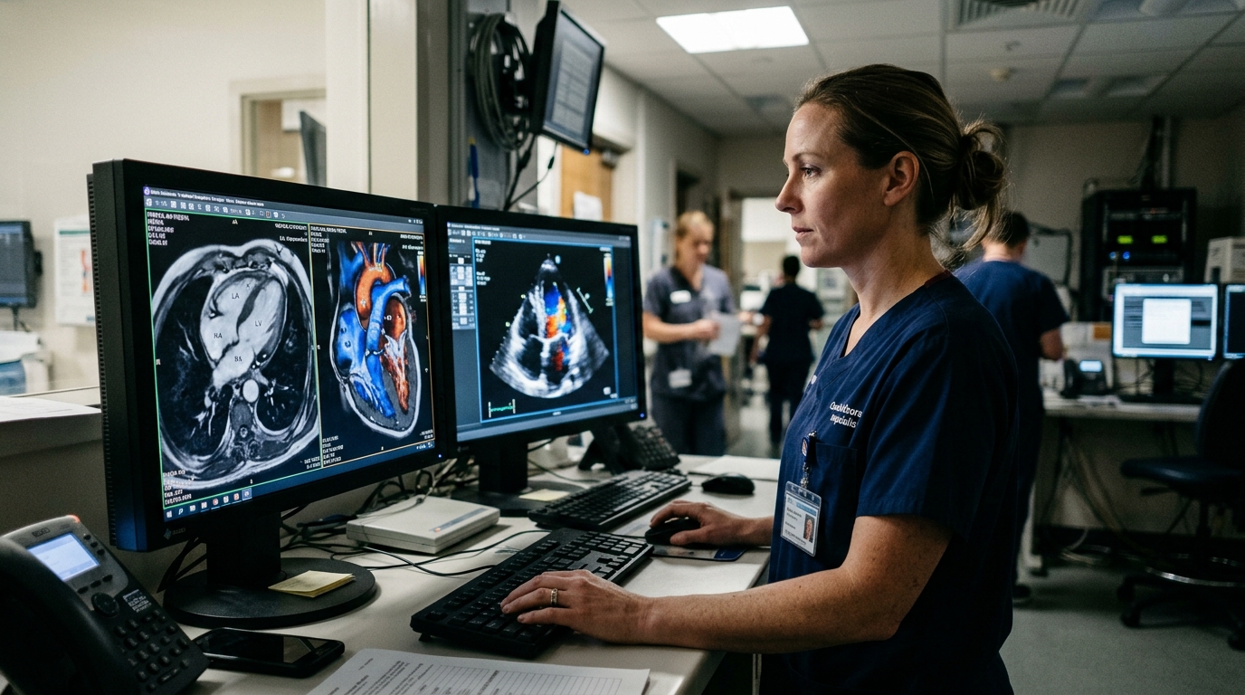
Echocardiography sits at the center of this work-up. Transthoracic echocardiography provides rapid, non-invasive assessment of cardiac structure and global function, while transesophageal echocardiography offers superior visualization of the highest-risk structures, particularly the left atrial appendage, prosthetic valves, and the thoracic aorta. The review highlights how three-dimensional transesophageal imaging and multiplanar reconstruction have expanded these capabilities, improving evaluation of the left atrial appendage by delineating trabeculations, lobar anatomy, and thrombus morphology, sharpening the assessment of prosthetic valves, intracardiac masses, and endocarditic vegetations, and helping differentiate thrombus from imaging artifacts in borderline cases. Three-dimensional transesophageal echocardiography is also described as essential for planning and guiding structural interventions such as left atrial appendage occlusion, where accurate sizing and spatial orientation are critical.
When echocardiography cannot answer the question, the review assigns complementary roles to other modalities. Cardiac computed tomography offers very short acquisition times and high spatial resolution of roughly 0.4 to 0.6 millimeters, but its soft-tissue contrast is limited, it involves ionizing radiation, and it requires iodinated contrast agents with potential nephrotoxicity. Cardiac magnetic resonance provides superior tissue characterization and remains the reference standard for detecting left ventricular thrombus, myocardial scar, and distinguishing thrombus from tumor in selected cases. Both chest CT and CMR can visualize thoracic structures beyond the heart itself, identifying extracardiac sources of embolism such as malignant cardiac invasion or disease of the entire thoracic aorta. The authors stress that these tools should be individualized and reserved for scenarios in which echocardiographic assessment is insufficient.
To organize the diagnostic pathway, the review proposes three broad clinical scenarios. In elderly patients with multiple cardiovascular risk factors, priority goes to excluding large-artery atherosclerosis with carotid Doppler ultrasound and, when appropriate, CT angiography, and to investigating occult atrial fibrillation with prolonged rhythm monitoring, with transesophageal echocardiography reserved for suspected high-risk substrates or inconclusive non-invasive imaging. In patients with signs of infection, such as fever, positive blood cultures, prosthetic valves, or intracardiac leads, infective endocarditis must be prioritized, prompting early targeted imaging for valvular vegetations or device-related infection. In younger patients without traditional vascular risk factors, the focus shifts toward congenital abnormalities, right-to-left shunts, and intracardiac masses, where transthoracic and transesophageal echocardiography are first-line tools.
Atrial fibrillation receives the most detailed treatment, since it is the most frequent cardiac source of cerebral embolism, with thrombi arising almost exclusively from the left atrial appendage. Transthoracic imaging gives an initial overview of atrial size and ventricular function, but targeted appendage assessment relies on transesophageal echocardiography, which remains the reference method for detecting appendage thrombus, spontaneous echo contrast, and markedly reduced emptying velocities, findings the review calls powerful indicators of stasis and thrombotic risk. Multilobulated appendages and non-chicken-wing morphologies have been associated with higher thromboembolism in observational cohorts, though morphology alone is insufficient for decision-making. Current European guidelines base anticoagulation decisions on the CHA₂DS₂-VA score, with oral anticoagulation indicated at a score of 2 or more and direct oral anticoagulants preferred in non-valvular atrial fibrillation, yet a residual cardioembolic stroke risk of approximately 15 percent persists despite appropriate anticoagulation. Percutaneous left atrial appendage closure holds a Class IIb recommendation for patients at high thromboembolic risk who cannot maintain long-term anticoagulation, while surgical appendage closure during concomitant cardiac surgery carries a Class I recommendation, though surgical closure alone cannot currently replace systemic anticoagulation because no randomized study has directly compared the two.
The prosthetic valve section underscores how imaging technology is reshaping the field. Mechanical prostheses carry thrombosis rates of 0.3 to 8 percent per patient-year, higher in the mitral and tricuspid positions, and cardiac CT has become the reference for detecting hypoattenuated leaflet thickening, a manifestation of subclinical leaflet thrombosis. In the PARTNER 3 CT substudy, subclinical leaflet thrombosis was identified in 23 percent of transcatheter aortic valves and 12 percent of surgical bioprostheses at one year, mostly asymptomatic and undetectable by echocardiography. A recent systematic review and meta-analysis of 176 patients undergoing transcatheter tricuspid valve replacement reported an overall thrombosis incidence of 15.8 events per 100 patient-years, with clinical thrombosis in 1.7 percent and subclinical leaflet thrombosis in 9.7 percent over a weighted mean follow-up of 10.2 months. The authors also report their own single-center experience with newest-generation transcatheter bioprostheses, noting a relatively high number of thrombosis events even under immediate postoperative heparin therapy, most clinically silent and marked mainly by modest gradient increases, and they call for larger systematic studies to define true incidence and optimal prevention.
Left ventricular thrombosis, historically linked to large anterior myocardial infarction, has markedly decreased with widespread primary percutaneous coronary intervention, but it persists in delayed-presentation STEMI and large infarctions, with contemporary CMR series finding an incidence of approximately 6.3 percent in unselected STEMI cohorts, rising to 12.2 percent in anterior STEMI and 19.2 percent when ejection fraction is below 50 percent. Transthoracic echocardiography remains first-line for detection despite limited sensitivity of 21 to 35 percent in post-infarction studies, improving to 70 percent in anterior STEMI with reduced ejection fraction, while contrast-enhanced imaging raises sensitivity to 61 to 64 percent and cardiac magnetic resonance serves as the reference standard. On prevention, the review notes that once left ventricular thrombus is diagnosed, oral anticoagulation should be considered for 3 to 6 months per current European guidelines, and that a randomized trial of low-dose rivaroxaban added to dual antiplatelet therapy significantly reduced thrombus formation after anterior STEMI without increasing major bleeding.
Congenital and structural causes complete the picture. Patent foramen ovale, present in roughly 25 percent of the general population and up to 50 percent of patients aged 60 or younger with cryptogenic ischemic stroke, is evaluated through a stepwise pathway beginning with contrast transthoracic echocardiography or transcranial Doppler for screening, followed by contrast transesophageal echocardiography for definitive anatomical assessment. Randomized trials including RESPECT, CLOSE, REDUCE, and DEFENSE-PFO have consistently shown that percutaneous closure reduces recurrent ischemic stroke compared with medical therapy alone in selected patients younger than 60 with cryptogenic stroke and high-risk anatomy, with the greatest benefit in large shunts or atrial septal aneurysm. Among cardiac masses, myxoma is the most common primary tumor in adults, with neurological manifestations in about 25 to 45 percent of cases, and papillary fibroelastoma fragments can embolize, making surgical excision the definitive treatment for both. The review concludes that tailoring the work-up to these clinical scenarios enhances diagnostic efficiency, reduces unnecessary testing, and supports timely identification of the true embolic mechanism, the foundation for targeted stroke prevention.
Subject of Research: Diagnostic imaging strategies for identifying cardiac sources of cardioembolic stroke and guiding prevention
Article Title: Cardioembolic sources and stroke prevention: a systematic review
Article References: Cozza, E., Cito, V., Giumbini, G., Marsili, G., Barletta, M., Zannoni, J., Mainardi, C., Bedogni, F., & Tusa, M. (2026). Cardioembolic sources and stroke prevention: a systematic review. Cardiovascular Ultrasound, 24(1), Article 8. https://doi.org/10.1186/s12947-026-00367-5
Image Credits: AI Generated
DOI: 10.1186/s12947-026-00367-5
Keywords: cardioembolic stroke, echocardiography, atrial fibrillation, left atrial appendage, prosthetic valve thrombosis, patent foramen ovale, left ventricular thrombus, infective endocarditis, cardiac CT, cardiac MRI, stroke prevention, Cardioembolic
News Source: Cassandra Pierce. (November 6, 2026). Cardiac imaging takes center stage in hunting stroke’s hidden embolic sources. Scienmag.