Pelvic organ prolapse, a common and progressive pelvic floor disorder in which the bladder, uterus or rectum descends into the vaginal space, has long been studied through separate molecular lenses: collagen loss, oxidative injury, hormonal decline and fibroblast dysfunction. A new narrative review published in Frontiers in Cell and Developmental Biology argues that these fragments belong on a single axis, in which chronic mechanical loading alters tissue mechanics, mechanotransduction pathways shift fibroblast behavior, and extracellular matrix imbalance becomes the structural endpoint of failing support. The authors, led by Zhenglin He of Chongqing Health Center for Women and Children and Lubin Liu of West China Hospital, Sichuan University, propose that the disorder emerges when repeated deformation outpaces the tissue’s capacity for adaptive repair.
The review, published on 30 October 2026, organizes the evidence into six mechanistic layers: a pathogenic setting of pregnancy, vaginal delivery, ageing, menopause and elevated intra-abdominal pressure; an upstream bridge of matrix stiffness, integrin sensing, cytoskeletal tension and TGF-beta and p38-linked signaling; a central effector layer of fibroblast activation and impaired repair; a bridge-amplifier layer of oxidative stress, autophagy disturbance, apoptosis and senescence; a structural endpoint of collagen imbalance, elastin disruption and MMP/TIMP dysregulation; and modifier layers of hormones, genetics, omics and immune remodeling. The authors stress that this is a narrative synthesis, not a systematic review or meta-analysis, and that each evidence source was weighed by the tissue or model examined, the intervention performed and the inference the data can actually support.
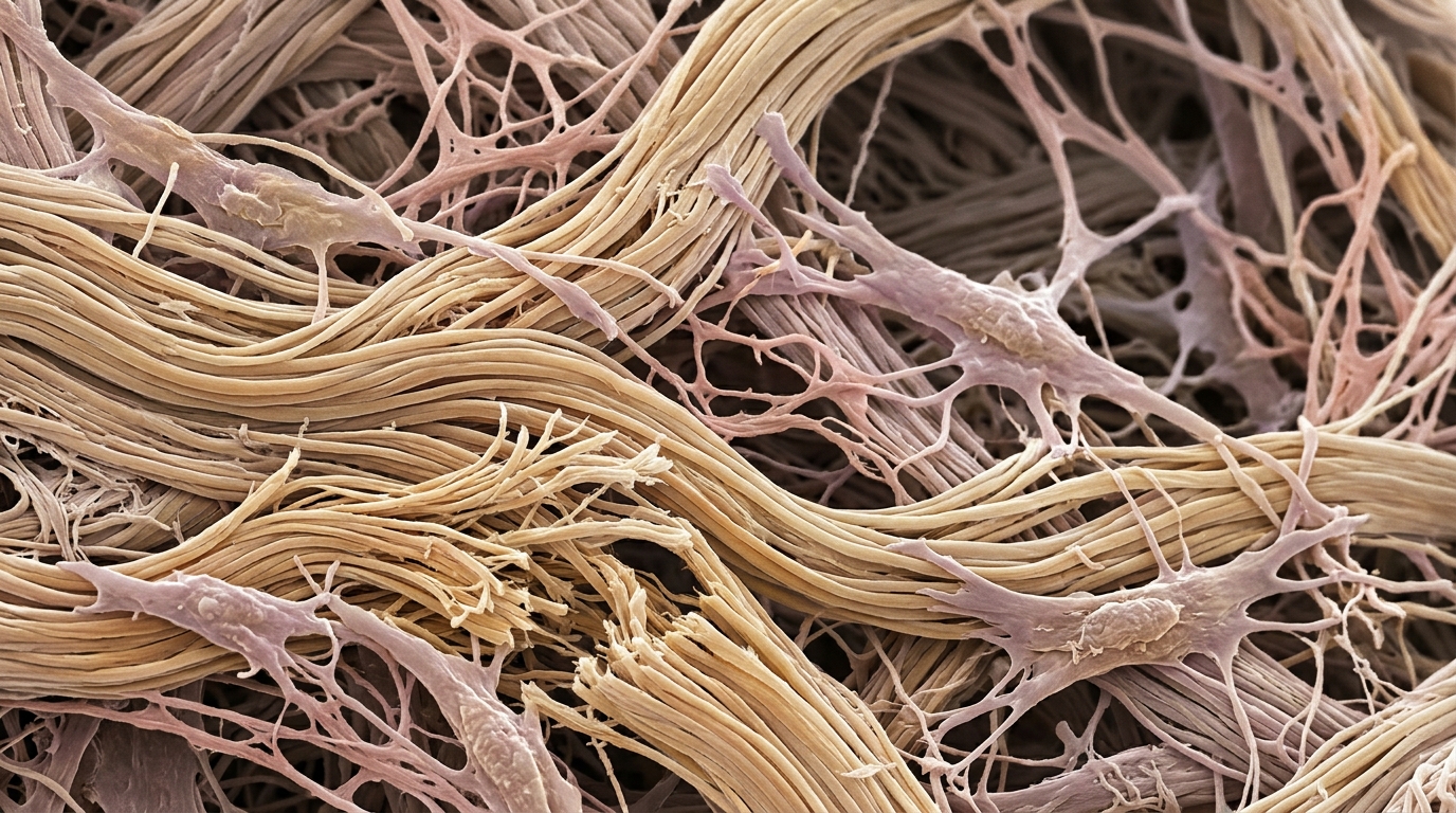
The mechanical starting point is distinctive. Vaginal delivery may damage levator ani muscles, fascial attachments and the vaginal wall, creating defects in a support system that then experiences recurrent loading. Changes in intra-abdominal pressure during coughing, lifting and defecation translate into tensile and shear forces across the vaginal wall and apical supports, and the extracellular matrix must sustain these cyclic, multidirectional loads while retaining compliance and recoil. Collagen fiber organization contributes tensile resistance, while FBLN5- and LOXL1-associated elastic-fiber homeostasis contributes recovery after deformation. Under physiological conditions, connective tissue remodeling and cellular repair can preserve integrity; the problem arises when loading becomes excessive, repetitive or prolonged and the reparative response no longer restores mechanical competence. Repeated loading becomes injurious when microdamage accumulates faster than the tissue can restore collagen architecture and elastic-fiber integrity, and the resulting structural changes redistribute strain, generating local mechanical cues to which resident cells respond.
Mechanically controlled experiments have begun to identify how loading regulates pelvic-support fibroblasts. In one study, primary vaginal fibroblasts from postmenopausal women with and without prolapse were exposed to uniaxial cyclic stretch at 0.1 Hz and 10% elongation; stretching altered the organization of cell-associated matrix, with disrupted networks observed in the prolapse-derived cultures. Experiments combining mechanical stress with hydrogen peroxide in uterosacral-ligament fibroblasts further linked physical and oxidative inputs to TGF-β1-associated MMP/TIMP dysregulation. Matrix rigidity provides a second, distinct experimental input: substrate-composition and stiffness studies using vaginal fibroblasts have linked the mechanical environment to myofibroblast differentiation and ECM production. In stiffness-controlled L929 fibroblasts, autophagy-dependent redistribution of Kindlin-2 promoted MOB1 degradation and nuclear YAP signaling, and autophagy inhibition partially reversed the myofibroblast response. Increased cytoplasmic Kindlin-2 and nuclear YAP were also observed in prolapse tissues, but the perturbation experiments were performed in a fibroblast cell line rather than intact human pelvic-support tissue.
Fibroblasts are among the most abundant stromal cells in pelvic supportive connective tissues, responsible for maintaining collagen turnover, elastin integrity and broader matrix homeostasis. Physiological activation supports matrix renewal after injury, whereas persistent myofibroblast differentiation can maintain contractility and abnormal matrix turnover. Prolapse-related fibroblast studies document altered collagen regulation, apoptosis and stiffness responses alongside increased matrix degradation in human support tissues. The review emphasizes that abnormal repair concerns matrix composition, organization and durability, not simply the amount of collagen produced. Fibroblast activation in prolapse shares features with fibrosis, including myofibroblast differentiation, TGF-β/p38-related signaling, increased contractility and stiffness-sensitive matrix production. Yet pooled human studies show reduced type I collagen, increased type III collagen and elevated MMP-1, MMP-2 and MMP-9 activity alongside elastic-fiber abnormalities, so fibrosis-like remodeling is part of the pathobiology but excessive collagen deposition does not adequately define the clinical disease.
Stress-intensifying processes deepen the repair failure. In cultured uterosacral-ligament fibroblasts, hydrogen peroxide altered collagen metabolism and the MMP/TIMP balance, and combined mechanical and oxidative exposure implicated TGF-β1-associated signaling. More directly, ATF3 silencing reduced hydrogen-peroxide-associated ROS, apoptosis, MMP2 and MMP9 expression and extracellular matrix loss through p38/NRF2-related signaling in vaginal fibroblasts, while SGK1-dependent regulation of GSK-3β/Fyn/NRF2 limited oxidative injury and matrix degradation in human uterosacral-ligament fibroblast models. Autophagy acts in a context-dependent manner: elevated miR-19-3p in vaginal fibroblasts was associated with reduced IGF-1 and COL I expression and increased autophagy and apoptosis through inhibition of AKT/mTOR/p70S6K signaling, whereas stiffness-induced autophagy promoted Kindlin-2 redistribution and myofibroblast conversion in L929 cells. The authors caution that neither finding justifies a general assumption that increasing or suppressing autophagy would benefit all prolapse tissues.
Apoptosis and senescence further constrain the repair capacity. Human prolapse tissue and fibroblast studies have implicated mitochondrial oxidative injury, while experimentally induced hypoxia and advanced glycation end-product exposure activated apoptosis-related pathways in uterosacral-ligament fibroblasts, and AGE–RAGE/MAPK/NF-κB signaling has been linked to reduced collagen I expression in vaginal fibroblasts. Senescence may sustain remodeling even when fibroblasts survive: prolapse-derived human uterosacral-ligament fibroblasts exhibited greater senescence and ROS burden together with lower SIRT1 expression, and pharmacological SIRT1 activation attenuated these phenotypes in culture. In that study, the senescent-cell fraction was 43.63% ± 10.62% in prolapse-derived cultures versus 4.84% ± 5.32% in control cultures. The authors note that secretory markers and acute injury models do not by themselves establish the temporal course of human prolapse.
Quantitative biomechanics now distinguish local matrix stiffness from organ-level support function. In fibroblast-generated extracellular matrices, the median effective Young’s modulus was 16 kilopascals for controls, 26 kPa for premenopausal prolapse and 50 kPa for postmenopausal prolapse, with five samples per group and a significant control-versus-postmenopausal difference at P = 0.0317; these values describe cell-derived matrices, not intact vaginal tissue. Planar biaxial testing of human vaginal tissue has demonstrated menopause-, stage- and direction-dependent moduli, including reduced toe-region moduli in postmenopausal stage-III prolapse relative to selected comparator groups. Mechanical competence also depends on recoil, extensibility and resistance to repetitive loading, so reporting only increased stiffness obscures the fatigue and multiaxial demands of pelvic support.
The translational section surveys repair strategies at very different maturity levels. An injectable fibrous hyaluronic-acid hydrogel used to augment uterosacral ligament suspension in rats improved the 24-week load at failure from 0.89 ± 0.28 newtons for suspension alone to 1.37 ± 0.31 N with hydrogel augmentation, while the intact ligament reached 1.70 ± 0.36 N; the authors note this provides a functional mechanical endpoint in an animal model but does not establish long-term clinical efficacy. Recombinant humanized collagen III increased proliferation, migration and ECM synthesis while reducing senescence in prolapse-derived vaginal fibroblasts, and in a rat simulated-birth-injury model was associated with higher collagen and angiogenesis-related markers. TNF-α-conditioned mesenchymal-stem-cell exosomes improved selected vaginal-wall ECM and inflammatory markers in rats with pelvic floor dysfunction, and puerarin-loaded electrospun patches combined sustained release with anti-inflammatory and pro-collagen effects, though their in vivo testing used a rat abdominal-wall defect rather than a prolapse repair model. Antioxidant and NRF2-related interventions have modified redox injury and MMP/TIMP regulation in pelvic fibroblast experiments, and SIRT1 activation reduced oxidative stress and senescence in cultured uterosacral-ligament fibroblasts, but such agents should be evaluated against tissue-quality and mechanical outcomes before being combined with reconstructive materials.
The review concludes that pelvic organ prolapse is best understood as a disorder of mechanically ineffective matrix remodeling in a pelvic support system exposed to repeated deformation, in which fibrosis-associated fibroblast activity can coexist with collagen imbalance, elastic-fiber failure and increased matrix degradation. The authors call for priority studies that pair anatomically matched tissue histology with tensile and shear testing, incorporate menopausal status and prolapse stage, compare molecular phenotypes using spatial or single-cell methods, and pursue longitudinal sampling around pelvic injury together with multiaxial organotypic models to establish how oxidative stress, fibroblast activation, senescence and matrix failure interact over time.
Subject of Research: Mechanistic framework linking mechanical loading, fibroblast dysfunction and extracellular matrix remodeling in pelvic organ prolapse
Article Title: Pelvic organ prolapse: emerging concepts in molecular mechanisms and tissue repair
Article References: He, Z., Tong, H., Liu, J., & Liu, L. (2026). Pelvic organ prolapse: emerging concepts in molecular mechanisms and tissue repair. Frontiers in Cell and Developmental Biology, 14, Article 1937362. https://doi.org/10.3389/fcell.2026.1937362
Image Credits: AI Generated
DOI: 10.3389/fcell.2026.1937362
Keywords: pelvic organ prolapse, fibroblasts, extracellular matrix, mechanotransduction, oxidative stress, senescence, collagen, pelvic floor, tissue repair, biomechanics, myofibroblast, SIRT1
News Source: Drew Townsend. (November 5, 2026). Scientists map how mechanical strain, fibroblast failure and matrix collapse drive pelvic organ prolapse. Scienmag.