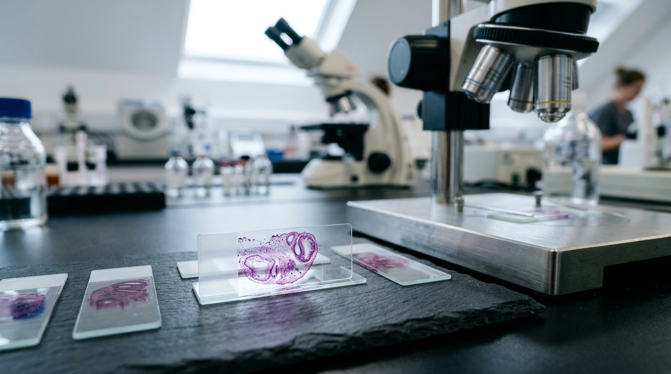
In an exploratory analysis of breast histopathology photomicrographs from a tertiary hospital in Nigeria, partitional clustering using K-means and Fuzzy C-means algorithms failed to recover inherited slide-level diagnostic labels separating benign from malignant tissue. While computer-aided diagnosis for breast histopathology has been developed almost exclusively using whole-slide images from high-income countries, many laboratories in sub-Saharan Africa operate with conventional light microscopes and camera-captured photomicrographs. Researchers investigated whether camera-captured images contain unsupervised structure corresponding to the benign–malignant distinction, but both clustering algorithms yielded two clusters characterized by weak internal structure and weak agreement with diagnostic labels.
The investigation analysed 14,084 tiles derived after quality control from 3,244 haematoxylin and eosin-stained photomicrographs of 186 patients, photographed at ×4, ×10, and ×40 magnifications. For each tile, researchers computed and standardised 118 handcrafted texture, morphological, and intensity descriptors. A swarm feature selection procedure run on 151 development patients, with 35 patients withheld, utilized an objective function combining the silhouette coefficient with patient-grouped cross-validated classification accuracy to retain 45 label-informed descriptors. Both K-means and Fuzzy C-means were subsequently evaluated across a sweep of two to fifteen clusters using internal validity indices and agreement with parent slide diagnoses measured by the adjusted Rand index.
Across the sweep of cluster counts, the silhouette coefficient peaked at two clusters for both algorithms, though internal structure remained weak. K-means clustering produced a silhouette coefficient of 0.2866, a Davies–Bouldin index of 1.3209, and a Calinski–Harabasz index of 5,759.30, while Fuzzy C-means produced a silhouette coefficient of 0.2794, a Davies–Bouldin index of 1.3704, a Calinski–Harabasz index of 5,676.43, and a partition coefficient of 0.6308. Agreement with the inherited slide-level diagnosis was minimal, yielding an adjusted Rand index of 0.0231 for K-means and 0.0226 for Fuzzy C-means. Both algorithms demonstrated a purity of 0.5806, which exactly equalled the majority-class baseline without offering any classification improvement.
Statistical evaluations revealed that cluster membership was more strongly associated with individual patient identity than with the diagnostic label, showing a normalised mutual information of 0.0419 for patient identity compared to 0.0109 for diagnosis, while association with image magnification was weak at 0.0073. Because technical factors were not measured in the study, this grouping cannot be conclusively attributed to technical rather than biological variation. The authors emphasized that these exploratory findings evaluate only a handcrafted, label-informed 45-descriptor representation and do not demonstrate that unsupervised methods in general cannot separate benign from malignant tissue, noting also that any quality-control application of clustering remains unvalidated.
Subject of Research: Unsupervised clustering of Nigerian breast histopathology photomicrographs
Article Title: Unsupervised separation of benign and malignant tissue in Nigerian breast histopathology photomicrographs: an exploratory evaluation of K-means and fuzzy C-means clustering
Article References: Odili, J. B., & John, O. (2026). Unsupervised separation of benign and malignant tissue in Nigerian breast histopathology photomicrographs: an exploratory evaluation of K-means and fuzzy C-means clustering. BMC Medical Imaging. https://doi.org/10.1186/s12880-026-03009-4
Image Credits: AI Generated
DOI: 10.1186/s12880-026-03009-4
Keywords: breast histopathology, unsupervised clustering, K-means, Fuzzy C-means, photomicrographs, computer-aided diagnosis, Nigeria, pathology, Unsupervised, separation, benign, malignant
News Source: Ophelia Keating. (November 5, 2026). Exploratory Analysis Finds Clustering Fails to Separate Nigerian Breast Histopathology Tissues. Scienmag.