


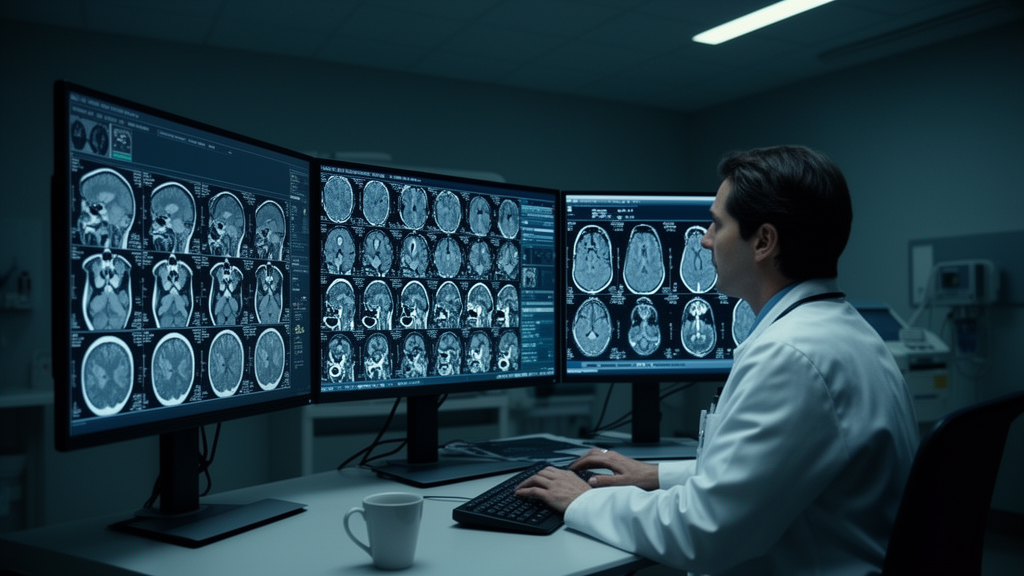
Two commercial, FDA-cleared artificial intelligence tools for detecting intracranial haemorrhage on emergency brain CT scans perform very differently when tested head to head, according to a retrospective diagnostic accuracy study published in BMC Medical Informatics and Decision Making. Analysing 3,884 consecutive non-contrast brain CT scans from adult emergency department patients at a single centre between 1 September 2024 and 28 February 2025, researchers found that one algorithm missed far fewer bleeds while the other raised far fewer false alarms, a trade-off the authors argue is crucial for hospitals deciding how to integrate AI into neuroradiological practice.
The study benchmarked each model’s output against the final neuroradiologist report, with every false-positive and false-negative finding adjudicated. Of the 3,884 scans, 421, or 10.8 percent, showed intracranial haemorrhage, comprising 183 intraparenchymal, 56 intraventricular, 201 subarachnoid, 183 subdural and 18 epidural bleeds. Both tools, the NeuroSuite CT ICH Assessment System (EFAI-ICH) and Rapid ICH (RAPIDAI-ICH), analysed 5 mm axial reconstructions of the same scans.
The high-sensitivity model, EFAI-ICH, identified 377 true positives against 44 false negatives, giving a sensitivity of 89.5 percent. RAPIDAI-ICH detected 308 true positives with 113 false negatives, a sensitivity of 73.2 percent. Specificity ran the other way: RAPIDAI-ICH reached 97.1 percent compared with 91.5 percent for EFAI-ICH, meaning the latter generated substantially more false alarms. Overall accuracy was higher for RAPIDAI-ICH at 94.5 percent versus 91.3 percent, and the specificity-favouring model also achieved the better positive predictive value at 75.7 percent, while EFAI-ICH delivered the superior negative predictive value at 98.6 percent.
In practical terms, the authors write, EFAI-ICH minimises missed bleeds but at the cost of more false alarms, whereas RAPIDAI-ICH excels at ruling out haemorrhage but risks under-calling subtle bleeds. Both platforms nonetheless demonstrated what the study describes as robust diagnostic capabilities for emergency ICH detection, with complementary strengths rather than one clear winner across all metrics.
The authors frame these performance variations as a starting point for deployment strategy rather than a definitive prescription. Their highlights raise the hypothesis that tiered deployment, or combined use of both AI tools, could be explored in future studies, since one model’s weakness is largely the other’s strength. Because the study was single-centre and retrospective, with performance measured against final neuroradiologist reports rather than a separate adjudicated gold standard independent of clinical practice, the reported trade-offs reflect one institution’s case mix and scanning protocol during a six-month window, and the findings are observational rather than evidence of improved patient outcomes.
The broader context the authors identify is that AI has become increasingly prominent in emergency neuroimaging, yet most clinical studies have evaluated only single diagnostic models, leaving comparative evidence on sensitivity-specificity trade-offs in AI-based ICH detection limited. This study’s contribution is precisely that comparison: two cleared commercial algorithms run on identical scans from the same consecutive patient series, with subgroup analysis by haemorrhage subtype. For emergency departments weighing which cleared tool to adopt, the results suggest the choice hinges on which error a service can better tolerate, missed subtle haemorrhages or excess false-positive alerts that add radiologist workload, and that recognising these variations is essential for optimal AI integration into neuroradiological practice.
Subject of Research: Comparative diagnostic accuracy of two FDA-cleared AI tools for detecting intracranial haemorrhage on emergency brain CT scans
Article Title: Unveiling diagnostic variability: clinical performance and pitfalls of two commercial AI tools for intracranial haemorrhage detection
Article References: Lee, S. Y., Chang, J., Wang, T.-H., & Chew, F. Y. (2026). Unveiling diagnostic variability: clinical performance and pitfalls of two commercial AI tools for intracranial haemorrhage detection. BMC Medical Informatics and Decision Making. https://doi.org/10.1186/s12911-026-03891-6
Image Credits: AI Generated
DOI: 10.1186/s12911-026-03891-6
Keywords: artificial intelligence, intracranial haemorrhage, CT imaging, diagnostic accuracy, emergency radiology, FDA-cleared algorithms, sensitivity, specificity, neuroradiology, retrospective study, Unveiling, diagnostic
News Source: Ophelia Keating. (October 30, 2026). Two FDA-Cleared AI Tools Show Opposite Trade-Offs in Detecting Brain Bleeds on CT. Scienmag.



