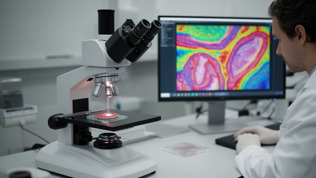
Researchers in Jena, Germany, have demonstrated that broadband coherent Raman scattering microscopy can distinguish tumor from healthy tissue in human head and neck cancer biopsies without any stains or labels, and they have paired that chemical specificity with an imaging setup roughly ten times faster than comparable multiplex CARS systems reported in the literature. The pilot study, published in Discover Imaging, combined tissue classification across five patients with a high-speed imaging demonstration on porcine skin and one human cancer sample, framing coherent Raman techniques as a candidate technology for ultrafast spectral histopathology.
The clinical motivation is straightforward. Current diagnosis of head and neck squamous cell carcinoma relies on hematoxylin and eosin stained biopsies interpreted manually by histopathologists, a process the authors describe as lacking the higher specificity urgently needed in oncology. Vibrational Raman microscopy can read the biochemical fingerprint of tissue non-invasively and label-free, but spontaneous Raman scattering has such a small cross section that pixel dwell times stretch to the order of a second, far too slow for practical imaging of large tissue sections.
Coherent Raman scattering techniques overcome that speed barrier. In coherent anti-Stokes Raman scattering, or CARS, two coincident laser pulses at pump and Stokes frequencies drive a vibrational coherence, generating an anti-Stokes signal through a four-wave mixing process. Stimulated Raman scattering, or SRS, instead detects the gain or loss of one beam after the interaction, a scheme inherently free of the non-resonant background that contaminates CARS and linearly proportional to oscillator concentration, which makes it attractive for quantitative analysis. Both nonlinear methods also co-generate second harmonic generation and two-photon excited fluorescence signals, enabling multimodal imaging that combines structural and chemical contrast without exogenous markers.
To reach the chemical specificity of spontaneous Raman, the team used broadband multiplex CARS, in which a broadband Stokes pulse with a narrowband pump probes the full Raman-active spectrum at once, requiring spectrally resolved detection. Their full-spectrum setup covered roughly 700 to 3200 cm−1, built on a 1032 nm CPA laser split into a spectrally narrowed pump and a white-light continuum Stokes beam, with stage scanning and a spectrograph coupled to a CCD camera. Five frozen tissue samples from five head and neck cancer patients were sectioned in parallel pairs: one stained with H&E and annotated by an experienced pathologist into labels including tumor, necrosis, dysplasia, inflammation, adipose tissue and connective tissue, the other measured with the broadband CARS setup and co-registered to those annotations.
Even a purely qualitative examination revealed strong spectral distinctions. Adipose tissue showed a prominent peak near 2845 cm−1 from CH2 stretching in lipids, while tumor regions displayed distinct signatures at 1653 and 1440 cm−1 and an increased signal between 1000 and 1200 cm−1, a range the authors attribute partly to elevated glycogen, which is typical of cancer-related structural changes. They caution, however, that this spectral window contains overlapping contributions from glycogen, lipids and proteins, so they interpret it as a composite signal rather than a glycogen-specific marker. Simple spectral projections, built by orthogonalizing average spectra of annotated regions with a Gram–Schmidt procedure, visually separated tumor from adipose tissue and even highlighted necrosis, tumor and connective tissue in samples from different patients, although spectral overlap between tumor and connective tissue remained evident.
For a quantitative test, the team applied principal component analysis followed by linear discriminant analysis, a statistical machine learning approach chosen deliberately for the small cohort. Spectra were preprocessed with interpolation, SNIP baseline correction and normalization using the RAMANMETRIX Python API, and models were evaluated with 10-fold cross-validation on a single field of view, optimizing the number of principal components between 1 and 30. The best configuration, using reconstructed full spectra with 15 micrometers of annotation erosion, reached a balanced accuracy of about 62 percent against a 25 percent random baseline for the four-class task. Removing the non-resonant background barely changed performance, suggesting the essential vibrational information is already encoded in the raw spectra. The authors stress this is far from clinical application, which would demand sensitivity and specificity approaching 100 percent, and they identify the small sample size, inter-patient variability and misalignment between annotations on parallel sections and the measured tissue as the main limitations.
The second half of the study tackled the speed problem directly. The custom high-speed setup pairs a commercial dual-output fiber laser providing a 791 nm narrowband pump and a broadband Stokes beam near 1030 nm with an ultrafast sCMOS camera and a multichannel lock-in detector, allowing simultaneous broadband CARS and SRS acquisition. The camera’s 29 microsecond exposure translates into a pixel dwell time of 134 microseconds, about an order of magnitude faster than typical multiplex CARS setups, which report dwell times of roughly 0.8 milliseconds for non-biological samples and 1 to 3.5 milliseconds for cells and tissues. The trade-off is spectral coverage: the fast system is limited to the CH-stretching region from 2800 to 3100 cm−1, and the authors note that a fair comparison across studies would also need to weigh signal-to-noise ratios, which cannot be assessed across different samples and laser configurations.
Demonstrations showed the speed is real on biological material. A 20-micrometer porcine skin section was imaged over a 2.5 by 0.25 square millimeter field of view, 4000 by 400 pixels, in 4 minutes and 24 seconds total acquisition time. Even with simple qualitative evaluation, the epidermis, dermis and subcutaneous adipocyte layers could be distinguished spectroscopically: the epidermis showed a stronger lipid-associated CH2 and CH3 stretching contribution, while the subcutaneous layer displayed the classic lipid signature with a strong 2850 cm−1 CH2 peak, a Fermi resonance near 2895 cm−1 and unsaturated fatty acid stretching around 3003 cm−1. The broadband CARS and SRS spectra agreed well in peak positions, with minor differences attributed to the techniques’ different concentration proportionality, detection schemes and spectral sampling. Applied to one human head and neck cancer sample, the setup captured a 1.9 by 2.5 square millimeter field at 3000 by 4000 pixels in under 50 minutes, reducible to about 30 minutes at a relaxed 1-micrometer pixel size.
The authors position these results as a proof of concept with clearly stated boundaries. The classification model was trained and validated on a single field of view from a five-patient cohort, annotation masks came from adjacent parallel sections rather than the measured tissue itself, and morphological erosion of the masks, while mitigating registration mismatch, inevitably obscures fine details such as cancer infiltration zones. Future work they envision includes larger, balanced multicenter datasets, H&E staining of the same sections used for imaging, deep learning models supported by adequate training data, and faster acquisition to make large-scale data collection feasible in the first place.
Still, the combination of chemically specific, label-free contrast with clinically relevant imaging speeds marks a concrete step toward spectral histopathology. With emerging laser sources and fast detectors, the authors expect ultrafast multiplex broadband coherent Raman imaging to extend into the Raman fingerprint region, the wavenumber range needed for tumor grading, staging and monitoring of compounds such as glycogen deposits. If that extension succeeds at maintained speed, the technique could eventually support real-time, label-free diagnostics where current standards are time-consuming and many diagnostic tools remain accessible only after surgery.
Subject of Research: Broadband coherent Raman microscopy for label-free classification and ultrafast imaging of head and neck cancer tissue
Article Title: Broadband coherent Raman microspectroscopy for the investigation of head and neck cancer advancing ultrafast spectral histopathology
Article References: Messerschmidt, C., Calvarese, M., Vafaeinezhad, M., Ryabchykov, O., Mühlig, A., Meyer-Zedler, T., Schmitt, M., Guntinas-Lichius, O., Bocklitz, T., & Popp, J. (2025). Broadband coherent Raman microspectroscopy for the investigation of head and neck cancer advancing ultrafast spectral histopathology. Discover Imaging, 2(1), Article 13. https://doi.org/10.1007/s44352-025-00017-y
Image Credits: AI Generated
DOI: 10.1007/s44352-025-00017-y
Keywords: coherent Raman scattering, CARS, SRS, head and neck cancer, spectral histopathology, label-free imaging, PCA-LDA, glycogen, porcine skin, biomedical optics, machine learning, tissue classification
News Source: Nathaniel Bowman. (October 30, 2026). Ultrafast Raman Imaging Speeds Toward Label-Free Head and Neck Cancer Histopathology. Scienmag.