


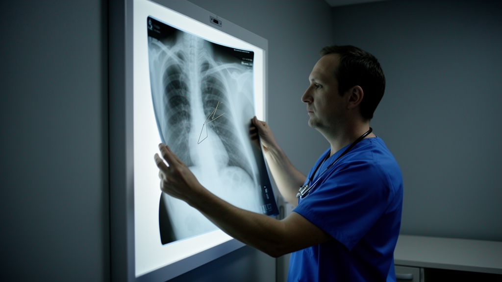
A temporary pacing wire that surgeons cut and left inside a patient’s chest after heart valve surgery fifteen years earlier migrated across the right side of his thorax and came to rest beside the airways and vessels of his middle lung lobe, where it was associated with collapsed lung tissue and coughing up blood. Surgeons in Japan removed the wire through small thoracoscopic ports using an intrapericardial route after dense adhesions made the usual pleural approach unsafe, and the patient recovered without complication.
Temporary epicardial pacing wires, known as TEPWs, are a routine part of cardiac surgery. They are sewn to the outer surface of the heart at the end of an operation so that the surgical team can pace the heart temporarily if the patient develops rhythm disturbances in the early postoperative period, and they are typically pulled out within a few days once the heart is stable. Occasionally, however, resistance is felt during removal, and the standard fallback is to cut the wire flush with the skin and leave the remainder in place. The authors of the report note that the long-term safety of this practice remains controversial, and that no standardized long-term follow-up protocol for retained wires has been established.
The patient in this report was an 80-year-old man who had undergone aortic valve replacement with a bioprosthetic valve, together with mitral valve repair for mitral regurgitation, fifteen years before his current presentation. He had severe aortic regurgitation and mitral regurgitation at the time, and a temporary epicardial pacing wire was placed during that operation. The operative record from the original surgery documented that resistance was encountered when the wire was pulled, so the surgeons transected it and intentionally left a portion in situ. For fifteen years the retained fragment caused no recognized problem, and its presence was apparently not tracked.
The wire resurfaced indirectly. During preoperative evaluation for cervical spondylotic myelopathy, the man underwent a chest computed tomography scan that incidentally revealed right middle lobe atelectasis. He was referred to the thoracic service for further workup. Bronchoscopy showed mucus obstructing the orifice of the right middle lobe bronchus and edematous narrowing of the distal mucosa, to the point that the distal bronchial lumen could not be visualized through the scope. Because malignant bronchial obstruction could not be excluded from these findings alone, the team initially adopted conservative management.
The situation changed when the patient developed hemoptysis, the coughing up of blood. Contrast-enhanced chest CT then demonstrated a continuous linear high-density structure approximately eleven centimeters long, extending from the right hilar region down to the inferior surface of the heart. The distal portion of this structure lay in the right hilum, adjacent to the middle lobe bronchovascular structures, and appeared to course near the inferior pulmonary vein. Importantly, the imaging showed no clear evidence that the wire had penetrated into the bronchial lumen, migrated inside the pulmonary vessels, or caused contrast extravasation or a pseudoaneurysm. Combining the imaging with his surgical history, the clinicians suspected that the dense line was the retained pacing wire and that its migration was associated with the atelectasis and the bleeding.
Because malignancy remained a possibility until tissue could be examined, the team performed a bronchoscopic biopsy of the abnormal bronchial mucosa on the morning of the removal operation. Frozen-section analysis showed no evidence of malignancy, clearing the way for the thoracic procedure. The surgeons began with a three-port video-assisted thoracic surgery approach, avoiding both a repeat sternotomy and a thoracotomy. What they found confirmed the difficulty of the case: dense adhesions around the hilum, particularly around the pulmonary veins, made dissection from the pleural side unsafe in that vascular territory.
The solution was to change anatomical planes. Working in collaboration with cardiac surgeons, the team opened the pericardium anterior to the phrenic nerve through the same thoracoscopic ports and dissected within the pericardial sac from the basal aspect of the heart toward the posterior pericardial space. The intrapericardial portion of the retained wire was identified near the inferior pulmonary vein and removed without bleeding or air leakage. No bronchial fistula, purulent fluid, or macroscopic evidence of infection was found, although bacterial culture of the extracted wire was not performed. The postoperative course was uneventful, and at eight-month follow-up the atelectasis had resolved on chest CT and the hemoptysis had not recurred.
The authors are careful about causation, and their caution is instructive. Direct bronchial penetration was never confirmed, so they propose that the bronchial edema and lung collapse may have arisen through indirect mechanisms, such as inflammatory bronchial narrowing, peribronchial irritation, or extrinsic compression related to a foreign-body reaction around the wire. Several observations were consistent with that interpretation: the absence of air leakage or bleeding after removal, the subsequent improvement of the atelectasis, and the lack of recurrent hemoptysis. The report also acknowledges the limits of the historical record. The original operative note did not document the exact placement site of the wire or whether the cardiac end had been suture-fixed to the epicardium, and no interval chest CT images existed between the index operation and the current presentation, so gradual migration over several years could not be excluded. The exact timing of extrapericardial migration could not be determined. Retrospective review showed the linear density was already present on the initial CT, while on plain chest radiographs the wire was difficult to identify because it overlapped the mediastinal and hilar structures.
Retained pacing wires are a recognized, if uncommon, late complication of cardiac surgery. The report’s summary table of previously published complications lists cardiovascular migration, intrathoracic migration into the chest wall or lung, and bronchial penetration, with removal approaches that have included transcatheter, transbronchial, and open thoracic techniques depending on where the wire ended up. What distinguishes this case is both the interval, fifteen years, and the location: a hilar, pericardial-adjacent position rather than definite intrabronchial or intravascular migration. Based on the CT course and the intraoperative findings, the authors judged the migrating end to be the transected cutaneous end, with the cardiac end presumed to have remained near the inferior cardiac surface, possibly on the inferior, or diaphragmatic, aspect of the right ventricle. Because the wire lay close to the pulmonary veins and hilar vessels, vascular injury remained a concern even without radiological signs of it, which is why cardiac surgeons were involved and why the intrapericardial route was chosen.
The practical lessons the authors draw are procedural rather than dramatic. When resistance is felt during wire removal, cutting the wire and leaving it behind is common practice, but this case adds to the evidence that retained fragments can migrate and cause symptoms many years later. They emphasize documentation of wire placement and removal attempts, notifying patients that a fragment has been retained, and keeping retained TEPWs on the differential diagnosis for unexplained thoracic, respiratory, infectious, or cardiovascular symptoms in any patient with prior cardiac surgery. For surgeons facing a retained wire near the hilum or pericardium, particularly when vascular injury is a concern or dense adhesions block the pleural approach, the intrapericardial technique demonstrated here may offer a controlled alternative. This is a single case report, so it cannot establish how often retained wires migrate or which removal strategy is best overall, but it documents a feasible solution to one of the harder anatomical variants of an old and largely forgotten piece of surgical hardware.
Subject of Research: Late intrathoracic migration of a retained temporary epicardial pacing wire causing atelectasis and hemoptysis 15 years after aortic valve replacement
Article Title: Late intrathoracic migration of a retained temporary epicardial pacing wire associated with middle lobe atelectasis and hemoptysis 15 years after aortic valve replacement: a case report
Article References: Kaneda, S., Miyahara, S., Mitsumata, S., Wakahara, J.-I., Midorikawa, K., Ueda, Y., & Sato, T. (2026). Late intrathoracic migration of a retained temporary epicardial pacing wire associated with middle lobe atelectasis and hemoptysis 15 years after aortic valve replacement: a case report. General Thoracic and Cardiovascular Surgery Cases, 5(1), Article 29. https://doi.org/10.1186/s44215-026-00274-1
Image Credits: AI Generated
DOI: 10.1186/s44215-026-00274-1
Keywords: temporary epicardial pacing wire, retained surgical wire, aortic valve replacement, hemoptysis, middle lobe atelectasis, thoracoscopic surgery, intrapericardial approach, chest computed tomography, bronchoscopy, cardiac surgery complication, foreign body migration, case report
News Source: Ophelia Keating. (October 29, 2026). Pacing wire left in the chest migrated 15 years after heart surgery. Scienmag.



